Warning: We observe an increase of emails from fake travel portals like . "travelhosting.co.uk". We never send links to such portals so be vigilant!

Neutrons for the Biotech Industry

Europe/Berlin
Facultad de Educación

Facultad de Educación

Campus San Francisco University of Zaragoza
Description

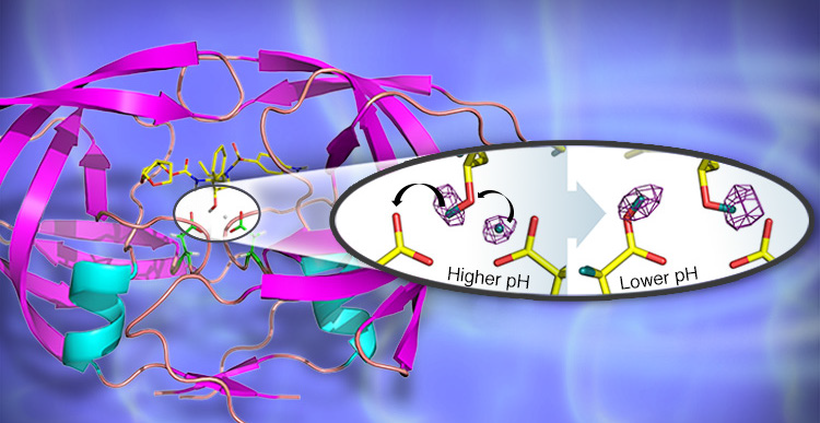
Neutrons are a non-destructive way of investigating biological and biotechnological materials. They can be used to study membranes, interfaces, surface morphology, solubility, macroscopic properties, crystal structure, colloids and much more. Neutrons can also locate individual hydrogen atoms on biological molecules and determine interactions between drug-molecules and their binding sites in the target. For more ideas of how neutrons can be used in biotechnology, please read our Case Studies.

Join us at the Aragon Materials Science Institute, ICMA (CSIC - University of Zaragoza) for SINE2020’s Neutrons for the Biotech Industry event where you can find out how neutrons can help your company and your research. At this event you will meet experts from the field of neutron scattering who will introduce you to several examples in their presentations. They will also be ready to discuss your particular questions in topic-related working groups. Learn also how to take advantage of our free proof-of-concept/feasibility studies via the SINE2020 programme.

Start: 13.00 Thursday 28th March 2019

End: 13.30 Friday 29th March 2019

Programme: Presentations and workshops on applications for neutron analytical techniques in the field of Biotechnology. More details coming soon.

Registration is FREE and the event also includes dinner on Thursday evening.

For more information about the SINE2020 project visit our website www.sine2020.eu

We are looking forward to welcoming you in March.

SINE2020 Industry Consultancy Team


Organising committee:

Javier Campo (ICMA)

Jesús Martínez de la Fuente (ICMA)

Germán Romeo (ICMA)

Mari Carmen Gámez (ICMA)

Lucy Moorcraft (TU Munich, GER)

Marc Thiry (Helmholtz-Zentrum Geesthacht, GER)


(Picture Credit: Oak Ridge National Laboratory)

  • Thursday, 28 March
    • 13:30 14:00
      Registration and Coffee 30m
    • 14:00 14:10
      Welcome 10m
      Speaker: Prof. Javier Campo (ICMA)
    • 14:10 14:30
      Introduction to SINE2020 20m
      Speaker: Marc Thiry (Helmholtz-Zentrum Geesthacht)
    • 14:30 15:00
      Neutron Scattering as a Versatile Tool to Study Structure and Dynamics of Proteins 30m

      The ability to respond to various environmental stimuli is a crucial ability of biological organisms. Light signal sensation and transduction, for instance, are important biological processes that allow organisms to respond to external stimuli. In recent work we have investigated structural and dynamical changes of a light-sensitive photoreceptor protein in response to light-illumination (1,2). In my presentation, I will present recent neutron spectroscopy and small-angle scattering experiments, and I will demonstrate how molecular dynamics and protein flexibility are required for light signal transduction of that photoreceptor. In particular, we could demonstrate that end regions of the photoreceptor protein change flexibility as response to light illumination. Hence, changes of protein flexibility are needed for light signal transduction of a biotechnologically relevant class of proteins. Neutron spectroscopy proved to be an excellent tool for that purpose.

      In the second part of my talk, I will present recent small-angle neutron scattering and reflectometry results from the field of biology and biophysics. A particular strength of neutron scattering is the concept of neutron contrast variation using a mixture of D2O/ H2O to render parts of the sample invisible to the neutron beam. This is a highly useful tool to selectively focus on specific structural aspects of a protein complex, to investigate crowding effects or highlight the specific interaction of proteins with biological membranes.

      In the last part of my talk I will speak about polymer-like properties of intrinsically disordered and unfolded proteins (3,4,5). By using neutron spin-echo spectroscopy combined with small-angle neutron scattering, we were able to study those flexible proteins. Polymer-like properties of those systems have been observed both in their structure as well as in their dynamics.

      Speaker: Andreas Stadler (FZ Jülich)
    • 15:00 15:30
      Neutron scattering of interpenetrating polymer networks (IPNs) as medical devices 30m

      A common material for urinary catheters is a hydrophobic polymer, silicone elastomer. The properties of silicone make it well-suited for producing medical devices; it has favourable mechanical properties and is chemically inert. However, this hydrophobic surface makes it prone to the adhesion of bacteria and subsequent and rapid formation of biofilms. The bacteria that grow in biofilms tend to be resistant to antibiotic treatment, and device-associated infections present a real challenge in modern medicine. To reduce the adhesion of bacteria and the risk of infection, the Danish company BioModics produces silicone catheters and medical devices that are functionalised by the inclusion of a hydrophilic hydrogel interpenetrating polymer network (IPN), which is introduced using supercritical carbon dioxide. This expands the silicone, and then hydrophilic monomers are introduced to react and form an IPN within the silicone network. This hydrogel not only reduces the risk of infection, but it is also has the potential to act as a drug delivery mechanism. The IPN acts as a reservoir for hydrophilic small molecules that can be suspended and then controllably released at site from the IPN-impregnated silicone.

      The release properties are dependent on the morphology of the IPN. However, it is a challenge to get insights to the hierarchical structure of the IPNs. As part of Denmark’s LINX Project (Linking Industry to Neutrons and X-rays), our team at the Niels Bohr Institute (University of Copenhagen, Denmark) performs neutron and X-ray scattering measurements on industrially-relevant materials working in conjunction with industry partners.

      A successful SINE2020 proposal was essential to determine the feasibility of using neutron scattering to obtain useful structural information about the IPN. We had already performed preliminary X-ray scattering measurements in the Niels Bohr Institute, but these only revealed the structure of the inorganic filler in the silicone and gave no information about the structure of the IPN component. In the SINE2020 experiments, samples of IPN were submerged in heavy water (D2O), which partitioned into the hydrophilic hydrogel and gave sufficient neutron scattering intensity to study the structure. Both small-angle neutron scattering (SANS) and spin-echo small-angle neutron scattering (SESANS) measurements were provided, covering a size range from ~1 nanometre to ~3.5 micrometre, which was important for a hierarchical structure such as these IPNs.

      These SINE2020 measurements have inspired further SANS and SESANS studies into more series of IPNs with different silicones and different concentrations and different hydrogels. I will describe how we initially assessed these materials using our in-house equipment (in the Niels Bohr Institute) and then by neutron scattering (via SINE2020) and have continued this since. I will then discuss these nanostructured materials as promising future medical devices and how the structure-property relationships arising from scattering measurements are assisting optimisation of the materials for the future.

      Speaker: Dr Gregory Smith (Niels Bohr Institute - University of Copenhagen)
    • 15:30 16:00
      Coffee Break 30m
    • 16:00 16:30
      Rational design of food processing methods with aid of neutron scattering 30m

      Systems of practical relevance to the food industry are often hard to investigate non-invasively. This is caused by the fact that most food emulsions are opaque and soft materials. The relevant length scales are often micrometres. Spin-echo small-angle scattering (SESANS) operates at these length scales and benefits from the high penetrating power of neutrons [1,2]. SESANS yields directly the scattering length density correlation function, which facilitates visual data interpretation [3].
      In the presentation the possibilities of SESANS will be illustrated with studies on the structure of protein gels [4,5], protein aggregates [2], protein mixtures [6], emulsions [1], colloids with tuneable interaction [7] and anistropic plant protein aggregates [8].

      References:

      [1] Effect of processing on droplet cluster structure in emulsion gels, A. Bot, F.P. Duval, and, W.G. Bouwman, Food Hydrocolloids 21 844–854 (2007)
      [2] Milk Gelation Studied with Small Angle Neutron Scattering Techniques and Monte Carlo Simulations, L.F. van Heijkamp, I.M. de Schepper, M. Strobl, R.H. Tromp, J.R. Heringa, W.G. Bouwman, J. Phys. Chem. A 114 2412-2426 (2010)
      [3] Real-space neutron scattering methods, W.G. Bouwman, J. Plomp, V.O. de Haan, W.H. Kraan, A.A. van Well, K. Habicht, T. Keller, M.T. Rekveldt, Nuclear Instruments and Methods in Physics Research A 586 9–14 (2008)
      [4] Characterizing length scales that determine the mechanical behavior of gels from crosslinked casein micelles, M. Nieuwland, W.G. Bouwman, M.L. Bennink, E. Silletti, H.H.J. de Jongh, Food Biophysics 10 416-427 (2015)
      [5] Relating water holding of ovalbumin gels to aggregate structure, M. Nieuwland, W.G. Bouwman, L. Pouvreau, A.H. Martin, H.H.J. de Jongh, Food Hydrocolloids 52 87-94 (2016)
      [6] Microstructure and rheology of globular protein gels in the presence of gelatin, Carsten Ersch, Marcel Meinders, W.G. Bouwman, M. Nieuwland, E. van der Linden, P. Venema, A.H. Martin, Food Hydrocolloids 55 34-46 (2016)
      [7] Direct comparison of SESANS and SAXS to measure colloidal interactions, K. van Gruijthuijsen, W.G. Bouwman, P. Schurtenberger and A. Stradner, EPL 106 28002 (2014)
      [8] On characterization of anisotropic plant protein structures, G.A. Krintiras, J. Göbel, W.G. Bouwman, A.J. van der Goot and G.D. Stefanidis, Food & Function 5 3233-3240 (2014)

      Speaker: Wim Bouwman (TU Delft)
    • 16:30 17:00
      NEUTRON SCATTERING – A VERSATILE TOOL TO BETTER UNDERSTAND PHOTOSYNTHETIC ADAPTATION, YOGURT FERMENTATION AND CHILI DELIVERY 30m

      Structural characterisation of biologically and biotechnologically relevant complex systems – using small-angle neutron scattering – is one of the research areas at the Neutronspectroscopy Department of the Wigner Research Centre for Physics and at the Budapest Neutron Centre. Here we demonstrate this through few selected examples.
      Our team has been investigating the structure and structural flexibility of photosynthetic membrane assemblies for many years. The range of samples extends from isolated thylakoid membranes through living algal cells to intact leaves. Monitoring the nature and extent of stress-induced membrane reorganisations is a key step towards the understanding the mechanism of stress responses in vivo. Small-angle neutron scattering (SANS), as a non-invasive technique, allowed us to reveal ultrastructural changes in different photosynthetic membranes under a large variety of abiotic and biotic stresses. Revealing the dynamic response of the system to illumination with varying intensity [1] and spectral composition [2] allowed us to better understand the photoprotective mechanisms in actions, while structural variations as a result of heavy metal ions or the trace elements [3] shed light to the influence of these pollutants on the photosynthetic organisms.
      SANS– a technique often applied to follow the sol-gel transformation of fermented dairy products – also helped us to characterize the influence of transglutaminase (mTG) on the fermentation process of bovine milk, and to demonstrate how mTG helps to retain the whey protein in yoghurt [4].
      Our group has also performed experiments on red chili pepper extract, which is used both as a food ingredient and a natural remedy for various medical conditions. Our scattering experiments provided information on the nanoscale pore characteristics of polyurethane microparticles, which can be used to entrap the extract in order to diminish its irritative potential [5].
      In the last example, I will discuss our recent results on high fat oil-in-water emulsions. With scattering measurements enabled us to characterize the interfacial structure of this system and the interaction between the applied emulsifiers sodium caseinate and phosphatidylcholine [6]
      References:
      [1] G. Nagy et al., Biochem J, 2011, 436, 225-30.
      [2] G. Nagy et al., Proc Natl Acad Sci USA, 2014, 111, 5042-7
      [3] O. Zsiros et al., Photosynth. Res., 2018
      [4] L. Darnay et al., Food Sci. Biotechnol 2015, 24, 2125-8.
      [5] L.-C. Borcan, Int. J. Nanomedicine, 2018, 13, 7155-66.
      [6] B. Yesiltas et al., submitted to J. Colloid. Interface. Sci.

      Speaker: Gergely Nagy (Paul Scherrer Institute)
    • 17:00 17:30
      Outside the box: Synchrotron Light for Biotechnology 30m

      Synchrotron facilities are a particular type of particle accelerator that produces synchrotron light, ranging from infrared up to X-rays, which is trillions of times brighter than conventional sources. Synchrotron Light is an extremely powerful tool for the characterization of a wide diversity of materials and processes at atomic level. The outstanding synchrotron light properties such as high brilliance, collimation and continuous spectrum, provides unique advantages compared to conventional techniques such as faster data collection, higher spatial resolution, lower detection limits or flexible sample environments to perform in-situ measurements.
      Synchrotron light applications to the health and biotech areas will be shown. These sectors are already taking profit from this valuable tool and there are many applications in these areas, as for example:
      • To study the interactions at atomic level of a target protein complexed with a new small molecule with therapeutic activity. It is extremely useful for the discovery of new drugs.
      • Characterization of pharmaceutical formulations and polymorphism studies.
      • Study the effect of a cosmetic product in the skin or hair, from the structural to the biochemical point of view.
      • Study the inner structure of a cell
      Very often, the industry and the academia are not aware of synchrotron techniques and their applications. For that reason, the European project CALIPSOplus brings together 14 synchrotrons and 8 FELs aiming at removing access barriers and promoting the usage of this type of facilities among the scientific and industrial community. In particular, TamaTA, a specific work package of CALIPSOplus, is granting SMEs access to this type of facilities.

      Speaker: Nuria Valls Vidal (ALBa Synchrotron)
    • 20:30 22:30
      Networking Dinner Restaurant

      Restaurant

    • 09:30 10:00
      Introduction to working groups
    • 10:00 11:00
      working group 1: Neutron scattering on polymer networks
    • 10:00 11:00
      working group 2: Industrial Access modes to Neutron Sources and Synchrotrons
    • 11:00 12:00
      working group 3: Structure and Dynamics of Proteins
    • 11:00 12:00
      working group 4: Synchrotron Light for Biotechnology
    • 12:00 13:00
      Summary and discussion
    • 13:00 14:00
      Lunch and Departure 1h