Choose timezone
Your profile timezone:
Warning: We observe an increase of emails from fake travel portals like . "travelhosting.co.uk". We never send links to such portals so be vigilant!

The SR2A – International Conference on Synchrotron Radiation and Neutrons in Art and Archaeology has been established since 2005 as the scientific event bringing together scientists and specialists from all over the world working on cultural heritage items with tools provided by large scale facilities. The conference offers an invaluable occasion for sharing cutting edge knowledge, expertise, experiences and new technical advances in the field of heritage sciences at large scale facilities.
In order to submit an abstract for SR2A 2023, please click on "Call for Abstracts" on the left. In order to register for the conference, please visit the registration website.
The aim of the conference is threefold:
1. to review the technological progress of advanced synchrotron and neutron research for the multiple research purposes on ancient materials, artworks and conservation methods
2. to foster the synergies between physics, natural sciences, humanities and social sciences, involving in particular preventive conservation and conservation science
3. to provide a forum for museum scientists, restorers, archaeologists, art historians etc. for bringing forward challenging issues in heritage science with advanced methods at large scale facilities.
After meetings, among others, in Amsterdam (2010), New York (2012), Paris (2014), Chicago (2016), Portsmouth (2018), a virtual meeting hosted by the Getty Conservation Institute (GCI) (2021), it is now to Munich to offer its appealing locations and institutions for this conference with world-class speakers.
The scientific topics cover among others: Archaeological advances, Paleontological and biomaterial advances, Ancient Materials, processes and production technologies, Technical advancements in synchrotron and neutron techniques, Multi-techniques and complementary methods for heritage sciences, Impact of analytical techniques on artefacts: non-destructive versus destructive analysis and the study of radiation-induced changes, Technical art history, Conservation science, Alteration processes and Monitoring of these processes.
Led by SR2A’s international committee, the 2023 edition will be organized in a cooperation with the Doerner Institut of the Bayerische Staatsgemäldesammlungen and the Heinz Maier-Leibnitz Zentrum (MLZ), a cooperation between the Technische Universität München (TUM) and two research centres of the Helmholtz Association, namely Forschungszentrum Jülich and Helmholtz-Zentrum Hereon.
Prof. Dr. Bernhard Maaz, General Director Bavarian State Painting Collections, Munich
Prof. Dr. Peter Müller-Buschbaum, Scientific Director FRM II / MLZ, Munich
Dr. Ina Reiche, Research Director CNRS, Paris, International advisory and program committee SR2A
Dr. Heike Stege, Doerner Institut, Munich, Local organizing team
In this presentation I will highlight the non-destructive techniques offered by neutrons providing unique information about archaeological objects on selected examples. Neutron tomography is largely complementary to X-ray CT with remarkably high penetration of metals and excellent sensitivity for organic materials. It has e.g. been used to elucidate manufacturing techniques used to produce ancient swords or helmets or to identify hidden artifacts within statues. Neutron-based analytical techniques have been used to analyze trace amounts of chlorine in the corrosion products of iron and bronze artefacts, while the elemental composition, especially the trace element contents is used in provenance investigations of ceramics, etc.
Advancing knowledge on the interactions occurring between conservation treatments and Cultural Heritage (CH) materials is a key point to plan effective conservation strategies. When dealing with inorganic-mineral products, the study of their diffusion within CH stone materials faces several analytical challenges, as non-destructive approaches, qualitative/quantitative phase analysis and high spatial resolution are simultaneously required.
Synchrotron radiation (SR) X-ray based 2D/3D/4D techniques have been recently used to successfully study the interaction of inorganic treatments, such as ammonium oxalate and ammonium phosphate (AmOx and DAP, respectively), with sedimentary stones of CH (i.e. micro X-ray powder diffraction in transmittance geometry (μ-TXRD) [2], micro-computed tomography (μCT) [2,3], micro-diffraction tomography (XRDCT) [4]). On the other side, a lot is still unknown on i) their reaction with artificial layered stone materials of CH as well as on ii) the crystallisation of newly-formed phases within or below the carbonatic painted layers.
This research shows the novel application of an advanced approach based on SR 2D X-ray based mapping techniques to characterize the effects induced by inorganic treatments (both AmOx and DAP) applied with different treatment modalities (poultice, capillarity) to painted plasters and XV century frescoes.
The high potential of SR high-lateral resolution 2D XRPD-XRF mapping has been applied to explore the: (i) AmOx and DAP reaction with Ca-rich and Mg-rich regions of carbonatic substrates; (ii) composition and localization of crystalline phases newly formed within treated substrates and their phase variation with the penetration depth; (iii) possible interactions of inorganic treatments with pigments and chromophore compounds. These elemental and structural investigations were carried out at the EBS-ESRF thanks to the new access route provided by the Historical Materials block allocation group (BAG) to the scientific community of CH [5].
The high quality datasets showed the crystallochemical composition and spatial distribution of phases of interest (down to single μ-particles of multiphase systems) within complex and heterogeneous matrixes. Moreover, the multi-analytical mapping approach highlighted the growth of a crystalline framework restoring the microstructural cohesion of painted layers and provided new knowledge on the variables affecting the reaction of inorganic treatments with carbonatic layered substrates.
Above all, this study allows a deeper understanding of consolidating mechanisms within CH geomaterials as well as opens novel analytical perspectives to explore the possible interactions of inorganic treatments with other crystalline and amorphous phases formed by decay processes and other/past conservation treatments.
References
[1] E. Possenti et al., Analytical Methods 12 (2020) 1587-1594
[2] E. Possenti et al., Materials Characterization 154 (2019) 315–324
[3] G. Massinelli et al., Construction and Building Materials 397 (2023) 132348
[4] E. Possenti et al. iScience 25 (2022) 105112
[5] M. Cotte et al., Molecules 27 (2022) 1997
Stone consolidation is a crucial practice in the conservation of building and stone artifacts of Cultural Heritage. To prevent the irreversible loss of stone material, with significant cultural value, various consolidants have been studied over the last decades. However, the diverse properties of stone materials (limestone, biocalcarenite, and sandstone), the state of conservation of the artifacts, and the use of diversified consolidants in the field have led to unresolved challenges in the consolidation practice. A comprehensive understanding of the consolidation process remains a significant analytical challenge, particularly regarding the effects induced by inorganic treatments (i.e., nano limes, ammonium oxalate, diammonium hydrogen phosphate) and the composition of major and minor crystalline phases formed within the treated stone materials (Sena da Fonseca, 2023).
This research is based on p an innovative approach using SR-µXRF and SR-µXRPD mapping carried out at the ID13 beamline of the European Synchrotron Radiation Facility (ESRF, Grenoble, France) to investigate the complex mixture of crystalline phases formed with ammonium oxalate solution (AmOx - (NH4)2C2O4) and diammonium hydrogen phosphate (DAP - (NH4)2HPO4)) on carbonatic porous stone substrates, whose phase identification is an analytical challenge (Osticioli, 2017). Here, the combined µXRF and µXRPD mapping approach has been used to study transversal cross-sections of AmOx and DAP-treated samples as well as double-treated ones (AmOx followed by DAP, DAP followed by AmOx). Noto limestone, a highly porous carbonatic stone widely used in the historical buildings of the Noto Valley (UNESCO's World Heritage List), was selected as a carbonate substrate. This synchrotron-based methodology thanks to its sub-micrometric resolution and high sensitivity (Cotte, 2022), resulted in an effective tool for studying the crystalline phases formed during consolidation improving our knowledge of the chemical and mineralogical transformations occurring within the stone matrix (Conti, 2023).
The PyMca ROI imaging software was employed as a tool to create average XRPD patterns over a selection of pixels and perform principal component analyses as well as batch-fit the XRF data (Cotte, 2016). The qualitative analysis of XRPD patterns of different regions of interest (ROI) was carried out. The analysis of distribution maps of specific crystalline phases with maps of marker elements via an RGB correlation enabled the identification of the crystal-chemical composition of the newly formed phases as well as the exploration of their spatial distribution at the microscale level, shedding light on the existence of stratigraphy of phases within the stone structure. The new calcium phases have been identified and localized in the calcite matrix; novel oxalate and phosphate phases have been observed in a mixture with phases detected in previous studies.
Moreover, this detailed analysis offered crucial insights into the phases' penetration depth. Additionally, it was possible to investigate the distribution of the new phases below the treated surface down to the inner portions of the stone matrix: this diffusion is essential to consolidate decayed portions and reconnect them with the sound original matrix. Furthermore, this methodology examines the crystallinity of these phases, and whether the newly formed phases exhibit preferential crystal growth. Understanding these aspects is crucial for optimizing consolidation treatments and promoting a homogeneous diffusion of treatments.
The experimental findings indicate that SR-µXRF and SR-µXRPD mapping is an efficient methodology to study the effects induced by inorganic-mineral conservation treatments for stone consolidation and gain a deeper understanding of consolidation mechanisms.
REFERENCES:
[1] Sena da Fonseca, B. (2023). Current Trends in Stone Consolidation Research: An Overview and Discussion. Buildings, 13(2), 403, https://doi.org/10.3390/buildings13020403.
[2] Osticioli, I., Botticelli, G., Matteini, P., Siano, S., Pini, R., & Matteini, M. (2017). Micro‐Raman analysis on the combined use of ammonium oxalate and ammonium phosphate for the consolidation and protection of carbonate stone artifacts. Journal of Raman Spectroscopy, 48(7), 966-971, https://doi.org/10.1002/jrs.5150.
[3] Cotte, M., Gonzalez, V., Vanmeert, F., Monico, L., Dejoie, C., Burghammer, M., ... & Susini, J. (2022). The “historical materials BAG”: A new facilitated access to synchrotron X-ray diffraction analyses for cultural heritage materials at the European synchrotron radiation facility. Molecules, 27(6), 1997, https://doi.org/10.3390/molecules27061997.
[4] Conti, C., Cutard, L., Botteon, A., Brambilla, L., Marinoni, N., Realini, M., ... & Colombo, C. (2023). Investigation of Calcium and Magnesium Phosphate Crystals in Stones Treated with Diammonium Hydrogen Phosphate Conservation Product: Potential of Micro-Raman Spectroscopy. Crystals, 13(8), 1212, https://doi.org/10.3390/cryst13081212.
[5] Cotte, M., Fabris, T., Agostini, G., Motta Meira, D., De Viguerie, L., & Solé, V. A. (2016). Watching kinetic studies as chemical maps using open-source software. Analytical chemistry, 88(12), 6154-6160, https://doi.org/10.1021/acs.analchem.5b04819.
KEYWORDS: Stone Consolidation, Ammonium Oxalate, Diammonium Hydrogen Phosphate, µXRF, µXRPD Mapping.
Museum objects with a painted metal structure are often found within industrial, scientific and technical collections. The conservation of these objects presents a challenge because of their composite nature. The interactions between the components of the system can lead to different alteration phenomena than what would be expected for the paint or metal alone, resulting in the need for specific conservation strategies. The CoPaiM project (Conservation of Painted Metals) seeks to contribute to the development of new conservation strategies for historic painted metal objects. A first research aim of this project is to enhance the knowledge of the material system and its alteration phenomena through the study of historic objects.
A corpus of painted iron objects from the 19th-early 20th century was studied using classic laboratory and synchrotron analytical techniques. Samples of paint and corrosion products were obtained from the objects and prepared as cross-sections. Synchrotron-based µ-XRF mapping was combined with µ-XANES point analysis to obtain spatially resolved information about the elemental composition and speciation of the components within the stratigraphy. This approach was complementary to the results acquired by optical microscopy and µ-Raman spectroscopy, and led to the detailed characterization of the paint components and iron corrosion products present in the samples and to a qualitative micro-scale description of the alteration phenomena. This study provides not only an improved understanding of the historical techniques used for the preparation of the objects, but also a documentation of possible materials and alteration states present in the painted metal system, the discernment of which is crucial for the development of suitable conservation strategies.
X-ray beams produced by a synchrotron source have properties of high brilliance and spatial coherence that make them highly suitable for studying a range of ancient materials. For example, synchrotron X-ray methods have been used to study fossil teeth to determine the age at death of humans using X-ray micro-tomography [1] and to identify chemical elements as markers of provenance from prehistoric sites and markers of diagenesis of Palaeolithic mammoth ivory using X-ray fluorescence [2]. However, high-flux X-ray measurements can induce chemical changes that can be visible to the naked eye (e.g. darkening of teeth [3]) or invisible (e.g. degradation of ancient DNA measured during quantitative polymerase chain reaction amplification [4]). The parameters governing the chemical mechanism and kinetics involved require further investigation.
In this work, we studied a corpus of fossil teeth (five specimens; 20,000 to 70 million years old) from five distinct animals and monitored changes using full-field multispectral X-ray excited optical luminescence (XEOL) imaging on the PUMA beamline of SOLEIL. After irradiation, evolution with time of the observed changes induced by the beam using photoluminescence (PL) micro-imaging was monitored.
During irradiation, we characterized the decay of the XEOL signal on a minute time scale under the high-flux X-ray irradiation as a function of the absorbed dose, the type of fossilized biological tissue (enamel, dentine), the incident energy, the detection conditions and the use of concomitant UV illumination. In particular, we found that UV illumination during and after the irradiation led to a faster and more complete recovery of the XEOL signal. On the enamel of a Mesohippus tooth, PL imaging after cessation of irradiation showed the presence of non-emissive lines correlated with the X-ray irradiation (immediately and 6 months after irradiation; Figure 1); however, no changes were observed in other tissues. The effect of the parameters on the decay of XEOL is being further investigated.
We have therefore identified characterization strategies to optimize the study of the secondary effects of irradiation and to better mitigate them in future experiments. This work is part of the general development of strategies to monitor, mitigate and better visualize the side effects of high-flux irradiation [5,6].
[1] P. Tafforeau & T. M. Smith (2008). Journal of human evolution, 54.2, 2008, 272-278.
[2] L. Tranchant et al., Quaternary International, 660, 2023, 4-12.
[3] G.D. Richards et al., American Journal of Physical Anthropology 149, 2012, 172-180.
[4] A. Immel et al., Scientific Reports 6, 2016, 32969.
[5] L. Bertrand et al., TrAC-Trends in Analytical Chemistry, 66.3, 2014, 128.
[6] L. Bertrand et al., TrAC-Trends in Analytical Chemistry 164, 2023, 117078.
This talk focuses on the urgent problems of synchrotron radiation damage to ancient paingtings. By the combined time resolved techniques of synchrotron radiation IR and ED-XAS in D-Line at SSRF, in situ radiation damage effect of paintings has been investigated. Results show that organic binders are easy to be damaged,especially when mixed with inorganic mineral pigments. And if the incident X-ray energy is near the absorption edge of major element in the pigments, the damage effect will be more serious. X-ray radiation damage effect will be more obvious from the point of view of protein structures, especially from their secondary structrues. This research indicates that, in analysis of ancient paintings by synchrotron radiation X-ray methods, radiation damage can be effectively reduced by using X-ray energy far away from the absorption edge of major element in pigments, or using time resolved techniques. These results have important reference value for how to reduce the radiation damage effect of cultural relic painting in synchrotron radiation X-ray analysis.
In the surroundings of the Pinakothek der Moderne
Ochre and related mineral pigments offer a fascinating insight into our deep past to examine cultural exchange, production methods and technical approaches. Analysis of Indigenous Australian ochre pigments on a variety of cultural materials such as boomerangs, bark paintings and rock art, reveals its composition, structure, and provenance. Recent archaeological science research at the University of Melbourne Archaeological Science Laboratory in collaboration with partners includes novel analysis of natural mineral pigments that utilize synchrotron methods to non-destructively examine provenance and composition (X-ray fluorescence microscopy), characterize chemical properties of unique organic binders (X-ray Raman and complementary laboratory-based techniques), and probe the changes to ochre due to cultural heating (synchrotron powder X-ray diffraction).
Determination of chemical composition can be a useful tool in provenance research of archaeological finds. The two fundamental levels of archaeometric investigation are the material characterization (i.e., the type of the matter) and the provenance identification (i.e., the source of the matter). Polished stone artefacts are especially appropriate subjects for the provenance approach since their chemical-mineralogical composition has usually not altered during their lifetime. By comparing the petrological-chemical features of archaeological objects with those of the potential raw materials, we can find how the archaeological items correlate directly with the raw materials and indirectly with the potential geological sources. Obviously, when investigating precious objects of cultural heritage, non-destructive methods are strongly preferred.
Prompt-gamma activation analysis (PGAA) is a non-destructive method for the determination of the bulk elemental composition (mostly major, a few minor and some trace elements) of different archaeological materials. The comprehensive research of polished stone tools has been performed since the 1990s, aimed of mapping the raw material procurement and the circulation as archaeological items in Hungary and surroundings from the Neolithic to Early Bronze Age (e.g. Szakmány 1996, Starnini & Szakmány 1998, Szakmány & Kasztovszky 2004, Szakmány et al. 2011, Bendő et al. 2019, Váczi et al. 2019, Szilágyi et al. 2021, Kereskényi et al. 2020, 2023). On the material characterization’s level, the main discriminative chemical constituents proved to be in general some of the major and minor elements (Si, Ti, Al, Fe and Mg) by applying Principal Component Analysis (PCA) on the PGAA dataset. On the provenance identification's level, major elements of alkali metals, Al, Fe and Ti could give further clue. Depending on the occurring rock types, raw material characterization and provenance determination can be done with medium-to-high certainty with the help of PGAA. Although PGAA proved to be a powerful non-destructive method to identify serpentinite, ‘white stone’ and hornfels, for other rock types it has limited success and requires complementary – sometimes destructive – methods (e.g. magnetic susceptibility, microscopic petrography, scanning electron microscopy).
The varieties of ‘greenish metamorphic rocks’ (greenschists, contact metabasites, amphibolites, blueschists, nephrites, high-pressure metaophiolites) can be characterised with similar chemical compositions, but with moderate differences in Ti, K, Mg, Mn and Fe. For example, blueschist implements proved to be originated from the Meliatic unit (Slovakia), while amphibolites are from the Gemericum-Veporicum (Slovakia). Five groups of nephrite were possible to be distinguished among the artefacts discovered in Hungary, most of them can be originated from the Lower Silesian Jordanów (Poland), while the origins of other four groups are uncertain (probably Alpine sources). High-pressure metaophiolites can be connected to the Alpine type eclogite facies metamorphites (Italy). Chemical overlapping of the ‘greenish metamorphic rocks’ can be found with magmatic rocks. The tools made of basic magmatic (or their slightly metamorphic varieties) rocks (basalts, metadolerites) can be connected to larger regional raw material units (SW Hungarian basaltic rocks of Mecsek Mts., NW Hungarian basalt types of Balaton-Highland, Kisalföld and the Novohrad-Gemer (Nógrád-Gömör) area) without exact raw material sources, mostly based on their elevated Ti and Fe content. Dolerite-microgabbro-metadolerite-metamicrogabbro lithic tools can be potentially connected to the Szarvaskő and Maros-valley geological sources. Serpentinites are Mg-Si-rich lithotypes with variable Cr or Ni content. Hornfels artefacts can be separated from other rock types by their Ca, Si, Ti and Fe content, and their provenance is related to the Maros-valley. ‘White stone’ is a term for a rock type with diverse but mostly Mg-Si- and/or Ca-rich composition. Serpentinites, nephrites, hornfels and Mg-Si-rich subgroup of ‘white stones’ comprise clearly distinguishable clusters.
Bendő et al. 2019. Archaeological and Anthropological Sciences 11: 1643–1667.
Kereskényi et al. 2020. Journal of Archaeological Science: Reports 32: 102437.
Kereskényi et al. 2023. Archeometriai Műhely XX/1: 1–22.
Starnini & Szakmány 1998. Acta Archaeologica Academiae Scientarium Hungaricae 50: 279-342.
Szakmány 1996. In: Makkay et al. (eds.) Excavations at Bicske-Galagonyás (part III). The Notenkopf and Sopot-Bicske cultural phases. Società per la Preistoria e Protostoria della Regione Friuli-Venezia Giulia, Quaderno 6. Trieste: 224-241.
Szakmány & Kasztovszky 2004. European Journal of Mineralogy 16/2: 285–295.
Szakmány et al. 2011. European Journal of Mineralogy 23: 883–893.
Szilágyi et al. 2021. Archeometriai Műhely XVIII/3: 237–260.
Váczi et al. 2019. European Journal of Mineralogy 31/5-6: 905–917.
Provenance research, i.e. identification of possible raw material sources of various archaeological objects, preferably using non-destructive methods, is a major task in Heritage Science. Prompt-gamma activation analysis (PGAA) turned out to be successful in provenance research of obsidians. Since the early 2000s, a significant database has been built at the Budapest Neutron Centre, which includes compositional data of about 500 geological and archaeological obsidian items, representing the major European and Mediterranean sources.
Besides the straightforward cases of provenance studies, however, there are still a few difficult questions to answer. During the excavations of a unique grave at Csongrád (SE Hungary), a long transparent obsidian blade, was found together with other objects. The grave was dated to 4370–4239 (1σ, 68.2%) cal. BC, belonging to the first wave of the ‘Pit Grave’ culture and having strong eastern contacts.
In a 1983 study by energy dispersive X-ray spectroscopy (EDS), the blade was compared to the Carpathian 1 (Slovakian) type raw material. In 2019, PGAA and portable X-Ray Fluorescence analysis have been done on the piece. Based on the first results, the Csongrád specimen, unlike other archaeological samples studied so far, was not possible to unambiguously associate with any of the known obsidian types.
As a further step, we have extended our database with more geological reference data from both sides of the Caucasus (i.e. from Armenia and Georgia), which are the closest outcrops to the main distribution region of the Pit Grave Culture in the NE part of the Black Sea. Until now, still no reassuring explanation is available concerning the provenance of the Csongrád blade.
Multiple bimetallic split-ear pommel swords from Northwestern Iran dated to the Iron Age (ca. 1250-800 BC) were investigated with Neutron Tomography at ISIS Neutron and Muon Source, UK. It is the first time that Iranian swords from that period were investigated with neutron techniques. The weapons were seized as part of law enforcement investigations and are pending repatriation. These circumstances, and the fact that there are many known examples of bimetallic weapons of this type being modified in modern times, led to a suspicion that a few were pastiches. With the use of Neutron Tomography, various modifications were revealed, including soldering and the use of other modern materials. Investigating bimetallic weapons is crucial to understanding the use of bronze and iron in the transitional period in early Iron Age Iran. Iranian weaponry, frequently found in museums but rarely well contextualised, is analysed in this study using non-destructive techniques to gain insight into ancient technology and modern forgeries.
More than 200 masterpieces of Venetian Painting of the 15th and 16th century are currently subject of a long-term cataloging and research project at the Bavarian State Painting Collections, Munich. In the Venice of the High Renaissance, a revolutionary change in painting style took place that had a decisive influence on the further development of European painting. The groundbreaking artistic ideas of this period are closely intertwined with equally innovative upheavals in painting technique and choice of materials.
The detailed comparison of current art-historical research with the latest visual and painting-technological observations is therefore of crucial importance. Thus, around 50 paintings – among them works by Titian, Veronese and Tintoretto – undergo extensive interdisciplinary examination by a team of art historians, conservators and scientists using manifold microscopical, imaging and analytical techniques to unravel their specific working process. The lecture will introduce the project, discuss actual case studies, and present exciting new findings.
Synchrotron-based X-ray Powder Diffraction (SR-XRPD) is particularly suited for the study of historical pigments. This technique indeed enables to discriminate between the multiple inorganic products present in complex paint stratigraphies at the micrometric scale, but also to provide detailed information on their composition and microstructure. This talk will present SR-XRPD research conducted via complementary analytical configurations, aimed at revealing the ancient syntheses of inorganic pigments, and deciphering the potential alteration mechanisms active in historical pictorial formulations. The communication will also present the “Heritage Materials” BAG implemented at the ESRF, and the opportunities offered by such collaborative initiatives to the Heritage community.
In 2019, Operation Night Watch started at the Rijksmuseum in Amsterdam. A wide range of macro-, micro-, and nanoscale techniques were used to study this 17th-century masterpiece by Rembrandt van Rijn (1606-1669). One of Rembrandt’s most commonly used pigments in The Night Watch (1642) is smalt, a ground potash glass colored blue by cobalt (Co) ions.[1] In this synchrotron radiation-based study, we used a combination of nanoscale X-ray fluorescence (XRF) imaging and ptychography, both in tomographic mode[2], to visualize and assess samples from different paint mixtures containing smalt. The experiments were conducted at beamline P06, Petra III, DESY(Hamburg, Germany).
Three samples were studied, taken from Co-containing areas in the painting that have different tonalities and pigment composition (Figure 1a). Due to the irregular shape and variation in size of the smalt particles it is difficult to assess the amount of smalt in the paint samples based on 2D techniques such as light microscopy or SEM-EDX. The 3D investigation of the samples at high spatial resolution enabled us to count the smalt particles, as well as to study their shape and volume. The analysis of the spatial correlation of cobalt with other elements present in smalt (As, Ni, and Bi) provided information about the production process, which in turn allowed determination of whether Rembrandt had used different types of smalt in The Night Watch. Figure 1c shows the 3D distribution of Pb, Fe, Ca, Co, Cu, Ti, and K in one of the samples used to provide semi-quantitative information on the composition of the paint mixtures and the differences between the three smalt-containing samples. Ptychographic reconstruction enabled visualization of the entire paint sample, including the organic fraction and components containing only elements lighter than sulfur, such as glass (SiO2).
[1] L. Robinet, M. Spring, S. Pagès-Camagna, D. Vantelon, and N. Trcera, Anal. Chem. 83, 2011, 5145–5152.
[2] K.W. Bossers, et al., J. Am. Chem. Soc. 142, 2020, 3691−3695
Infrared spectroscopy has become a routine method for studying artistic materials and monitoring degradation processes. Most studies focus on the mid infrared spectral region (MIR 600-4000 cm-1), whilst the far infrared region (FIR, 50-600 cm-1) is often neglected. However, some studies have already highlighted the potential of analyses in the far-infrared region. It was shown that many pigments consisting of oxides and sulphides, which are not detectable by MIR, could be identified by FIR spectroscopy. In many other cases FIR provides additional information to MIR spectroscopy.
Whilst prior studies used conventional infrared light sources and, as far as the authors know, reference materials produced nowadays, in the presented research focus is on the use of synchrotron-based far-infrared spectroscopy to characterize pigments from the legacy of the two painters Arnold Boecklin (1827-1901) and Edvard Munch (1863-1944). Analysing painting materials from the artists’ possession excludes by-products and impurities formed in modern production methods and show the composition of the original 19th and 20th centuries pigments.
Synchrotron-based infrared spectroscopy experiments were conducted at the IRIS beamline of the BESSY II synchrotron facility. SR-FIR spectroscopy provides much higher signal intensity than conventional experiments. This results in a higher quality of the spectra and allows identification of low-intensity bands which may not be visible in a spectrum obtained by conventional spectroscopy. Measurements in the FIR region were carried out in transmission mode.
The presented research will be the base for future investigations using synchrotron FIR-microscopy, which will allow the investigation of small samples and cross-sections, taken from objects, and comparing contemporary refences with materials used on artworks.
Within this talk, the technique of Small- and Wide-Angle X-ray Scattering (SAXS/WAXS) will be introduced in the context of nanostructure characterization of heritage objects especially of written artefacts or paintings.
The object of interest of the presented study is a fifteenth-century heritage object namely a set of 65 Tibetan ritual cards (tsakali) from a private collection. The obtained results of the 2D scanning SAXS/WAXS measurements of selected cards performed at the SAXSMAT beamline at DESY will be shown. In particular, the ability to use the diffraction signal (WAXS) to identify pigments will be highlighted. The aim is to better understand the provenance, molecular composition as well as the making and deterioration processes of that collection. Especially, if the collection has been made by one artist, is it one collection or is it composed of several sub-collections? Are there hidden layers underneath the visible painting of the tsakali cards? These are only a few research questions that can be answered partially by the analysis of the X-ray scattering patterns.
The selected showcases will highlight the potential of using SAXS and WAXS methods in the structural analysis of heritage objects in the field of archaeology and art and should also foster further development of using X-ray-based scattering techniques in that research field complementary to the well-established X-ray fluorescence technique.
We present analysis of seven samples from early modern English wall paintings, most dating to 1550-1660, when decorative wall paintings were used to convey status and cultural knowledge (Fig. 1A, 1B). The selection of pigments has implications for the affordability, accessibility, and sociocultural significance of the paintings. Identification of pigments and degradation products is also important for interpretation of the original colours. In the early 1600s, synthetic blue verditer (Cu$_2$CO$_3$(OH)$_2$) emerged as an important new affordable blue pigment alongside widely used but precious natural azurite, but other copper blues and greens were also in use.
While most samples were identified as verditer or natural azurite, a subset did not fit either profile. In many, elemental analysis showed colocalisation of copper and chlorine; while in some cases it was straightforward to assign these particles as degradation products, in other cases they were so widespread that the possibility of intentional use of copper chlorides as pigments was considered (Fig. 1C).
Further analysis of the sample subset that were not conclusively characterised as verditer or azurite was then carried out as part of the Heritage BAG at ESRF. First, µ-PXRD mapping of seven historical samples, selected for their unusual morphology and/or high chlorine content, was carried out. Additional experiments are planned at ESRF to address the detection and formation of copper hydroxychlorides in verditer/rouaite paint samples using µ-XRF and XANES.
Using µ-PXRD, we identified specific mineral phases (Fig. 1D). Several samples contained copper hydroxychlorides in the absence of carbonates, supporting the use of atacamite and other polymorphs as pigments in the wall painter’s palette.
Most significantly, rouaite, a basic copper nitrate, was identified in one sample for the first time. The localisation of rouaite and a basic copper carbonate in the paint layer stratigraphy suggests that rouaite is susceptible to degradation in the presence of chlorine, which may be one explanation of its infrequent identification in historical samples. Ongoing work in our lab investigating historical synthetic procedures for blue verditer synthesis has identified rouaite as a major undesirable byproduct, and the discovery of it in a wall painting sample is important evidence for a specific synthetic procedure as well as the unreliability of verditer production during this period.
We have demonstrated the complexity of the palette used in medieval and early modern England and the difficulty of conclusively identifying pigments and degradation products by visual examination and elemental analysis alone. This attests to the importance of multimodal analytic procedures and the utility of synchrotron radiation in the study of cultural heritage samples.
Paleolithic cave representations are one of the oldest forms of art from modern humans, and as such it is important to understand the coloring matters used, usually black, red or yellow. The study of black Paleolithic coloring matter has been well developed, and analytical procedures have been established [1],[2]. Instead, reds are more complicated, as the coloring matter and the cave wall present a similar chemical composition [3]. Thus, here we study the red coloring matter with two main aims. On the one hand, we want to enhance the red Paleolithic pigments’ characterization with respect to the in situ analyses in a cave that could be undertaken with a portable instrument. On the other hand, we would like to find differentiation criteria between the composition of the walls and that of the coloring matter, which could also help us to improve the performances of in situ analyses.
Red coloring matter from Altamira, La Garma, and several other Paleolithic key cave sites in Northern Spain was analyzed with micro-X-ray fluorescence (μXRF) at the PUMA beamline, SOLEIL synchrotron. Such sensitive non-destructive methods allow us to study trace elements in samples in a way that is not possible in situ as of today (namely with portable XRF, pXRF). A semi-quantitative treatment of the data is being developed. This synchrotron study was further complemented with SEM-EDX analyses, which provide a more detailed information on the surface morphology and chemical composition of the sample, such as the presence of impurities, as well as information on the distribution of elements like iron.
The combination of these two techniques, supported by others, made it possible to find differentiation criteria distinguishing the calcitic cave wall and the coloring matter, as well as to obtain information on the creation of the prehistoric figures. This allowed us to compare the representations between
them, in the same cave but also different caves between them. Finally, we further improved the identification of the elements to be searched for during in situ analyses with a pXRF equipment, such as Ti, Cr, Mn, Cu, Zn, Rb, Sr or Zr.
References :
[1] Trosseau et al. JAAS 2021
[2] Reiche, I. et al submitted Archaeometry 2023
[3] Reiche, I., Tapia, J. et al accepted Archéosciences 2023
Wall painting is one of the most recognizable legacies from the Roman period. Though rather limited in their use, lead compounds were present in the elaboration of their pigments, such as sandyx, described by Pliny. In the inner and upper layers of Roman wall paintings from the domus Avinyó archaeological site in Barcelona, lead-phosphorus compounds, together with the degradation product plattnerite (PbO2) have been detected. This raises concerns about their provenance: are they original pigments, or degradation products of lead-based pigments?
To answer these questions, historical painting samples have been studied by synchrotron radiation-based micro X-ray diffraction (µSR-XRD) and micro X-ray fluorescence (µSR-XRF). The high brilliance of synchrotron radiation-based techniques has demonstrated remarkable efficacy not only in detecting compounds of interest, but also in elucidating their distribution across different pictorial layers of the wall paintings.
The results obtained by these techniques have been complemented by optical microscopy (OM), scanning electron microscopy with energy-dispersive X-ray spectrometry (SEM-EDX), Fourier-transform micro-infrared spectroscopy (µFTIR) and micro-Raman spectroscopy.
In the surroundings of the Pinakothek der Moderne
Posters can be discussed with the authors.
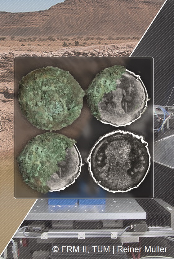
This work concerns the analytical phase of a project exploiting Synchrotron Radiation X-ray fluorescence (SR-XRF) spectrometry for the study of the composition of gold coins (Solidi) of the late Roman period. It is a project conducted at the Elettra Synchrotron in Trieste, on the study of the Treasure of Como: an exceptional discovery of 1000 late-imperial Roman coins (ca. 402-472 AD), found in 2018 in Northern Italy [1]. The research involves different professionals ranging from an archaeometric perspective that considers expertise from archaeology and numismatic to diagnostic, from physics and chemistry to science of materials.
The current study aims to investigate the composition and trace elements (Ag, Cu, Pt, Pd, Os, Ir, Ru, Rh, Sn, ...) of the coins issued in different mints and under different emperors, to obtain information on the raw materials and processes of making ancient gold. The sine qua non requirement was to adopt a totally non-invasive protocol, for this reason SR-XRF analysis was chosen to achieve the required sensitivity for trace element detection due to the high photon flux provided.
Also, a relevant aspect of this study was to use coins found in a well-defined archaeological context, as in the case of the Treasury of Como. The Treasury comes from an archaeological context known in its entirety and of which, thanks to studies already carried out, the mint of origin and date of issue are known. The context gives us more information about the association of coins, that is, it photographs the composition of the coins circulating at a given time, also allows us to date the concealment and to make proposals on the identity of those who have hidden the Treasury as well as making assumptions about the nature of the archaeological context.
The main challenge of the study discussed here is due to the high purity of Solidi. In fact, their high content of gold, close to 100% in weight, makes the trace elements hardly detectable. This, combined with the low penetration depth of X-rays in such a heavy matrix, have created an objective complexity in the quantitative determination of trace elements.
A recent XRF study on gold coins has adopted different approaches, such as subtracting pure gold reference spectrum from the spectra of each coin to solve signal overlapping issues [2] or using wavelength dispersion technique, allowing to achieve a spectral resolution capable of detecting trace elements [3]. In this context, the GOLD CoINS (GOLD Como INterdisciplinary Study) approach consisted of adopting reference materials with similar composition with coins or with variable content of elements of interest for defining the best measurement conditions in detecting trace elements. Three beam energies have been used to ensure a good detection of trace element-related signals useful to provide information on the compositional differences of coins and to enable the development of the next phase of statistical data analysis.
[1] G.Facchinetti, 2022, Il Tesoro di Como. Via Diaz 2018, Notiziario del Portale Numismatico dello Stato, 16.
[2] I.Carlomagno, P.Zeller, M.Amati, G.Aquilanti, E.Prenesti, G.Marussi, M.Crosera, G.Adami, 2022, Combining synchrotron radiation techniques for the analysis of gold coins from the Roman Empire, Scientific Reports, 12, 15919.
https://doi.org/10.1038/s41598-022-19682-8
[3] M.F.Guerra, E.Neri, M.Radtke, 2023, Gold leaf tesserae: tracing the origins of gold using synchrotron-based techniques, The European Physical Journal Plus, 138, 127.
https://doi.org/10.1140/epjp/s13360-022-03638-y
We have studied amphorae of the Haltern 70 type found in excavations of a Roman settlement at Castro do Vieito in the northwest of Portugal, as well as from the known kiln sites in Baetica and Lusitania, situated in nowadays Spain valleys of the Guadalquivir and Rio Tinto, and the coast near Cadiz and Algeciras, as well as Algarve Portugal coast.
Haltern 70 amphorae were produced mainly in the Roman provinces of Hispania Baetica and Lusitania in the south of modern Spain and Portugal, during the second half of the first century BC and the first century AC. These amphorae were used to transport agricultural products from the south to supply the Roman military stationed in the north.
During excavations of the Roman settlement at Castro do Vieito, a large number of such amphorae was found. The aim of our study is to find similarities in the element contents of sherds from the kiln sites and from Castro do Vieito that could prove that the amphorae from Castro do Vieito are from the south of Spain and possibly from which site they preferentially came.
By PGAA at FRM2 reactor at the Technical University of Munich, about 20 elements were determined from each sherd. The data have been analysed by principal component analysis and cluster analysis. The results show a clear clustering for many kiln sites, which is a necessity for provenance analysis. The unexpected result is that the finds from Castro do Vieito differ strongly from all sampled kiln sites. This suggests that the most probably kiln sites, the ones studied, are not the provenance of Castro do Vieito amphorae, or that there are still unknown production sites in the region.
In Ancient Egypt the lapis lazuli was the most precious gemstone as it was considered a sacred stone connected to the gods. Its special dark-blue colour symbolized the night sky and the starry heavens. Due to its symbolism and its scarcity, lapis lazuli had been used from the Predynastic period onward as the material of amulets, beads and inlays in jewellery. Despite its early presence and
some historical textual references, there is no physical evidence of a lapis lazuli mining area within the territory of Ancient Egypt. The most widely accepted concept is that the ancient Egyptian lapis lazuli originated from the Badakhshan Province (Afghanistan), however other new potential Asian extraction sites (Pakistan, Tajikistan, Lake Baikal area, Myanmar) were also suggested recently. This stone had reached Egypt most probably from Mesopotamia through trade routes, or could also be brought as a tribute from various localities like Assur, Babylon.
There are several ancient Egyptian artefacts of lapis lazuli in the Egyptian Collection in the Museum of Fine Arts, Budapest. In this project we aim to investigate these objects in order to determine the raw material and the provenance of their raw material.
The lapis lazuli amulets kept in the collection of the Department of Egyptian Art in the Museum of Fine Arts Budapest originate from the 19th century private collections. Their provenance is unknow. These amulets were made between the second and first millennium BC. The purpose of our examination was to establish the origin of the raw material used for the production of these amulets. In the focus of this examination were 23 different amulets, 5 djed-pillar amulets and 2 wedjat-eye amulets from the collection of the Department of Egyptian
Art of the Museum of Fine Art, Budapest.
As these artefacts are irreplaceable, we have to strictly apply non-destructive analyses.
Lapis lazuli is an opaque semi-precious stone consisting mainly of a blue mineral, lazurite, an alumo-silicate of the complex feldspathoid sodalite group, and can be described as an isomorphic combination of haüyn and sodalite. The chemical formula for lazurite is (Na,Ca)4(AlSiO4)3(SO4SCl), but with considerable variation in the amounts of SO4, S and Cl. Additionally, pyrite and calcite together with relatively small amounts of accessory minerals are present in the stone lapis lazuli.
Because lapis lazuli is very inhomogeneous, and the objects have often been exposed to the weathering for thousands of years, any surface measurements carry the risk of falsifying the results of the analysis. Prompt- gamma activation analysis (PGAA) is a nuclear analytical technique utilizes the characteristic prompt γ-ray spectrum measured during the irradiation of the sample for determination of elemental compositions.
Since neutrons can enter the deeper regions of the sample, they are ideal for bulk analysis. In the last years, several attempts have been made to apply PGAA, to determine the origin of lapis lazuli. The first results in distinguishing lapis lazuli raw materials of different occurrences (Afghanistan, Lake Baikal, Ural, Canada and Chile) were already published.
In addition, portable non-destructive techniques have been applied to the objects on-site at the Museum of Fine Arts, Budapest. In order to analyse the chemical composition pRFA (X-Ray Fluorescence Analysis) and in order to identify the mineralogical phases, Raman-Spectroscopy and Near-Infrared-Spectroscopy have been carried out. These techniques are similarly helpful, to identify lapis lazuli and other precious stones as neutron-based methods. The results obtained from complementary investigations have be combined with PGAA results.
The present work focuses on the archaeometric investigation of late-imperial Roman gold coins (solidi), issued by different imperial mints from pars Orientis and Occidentis and dated to about 402-472 CE. The examined coins are part of 1000 solidi of the “Treasure of Como”, a recent astounding discovery of precious and unique archaeological findings in Northern Italy [1].
The elemental fingerprint of archaeological coins can be related to the mints of origin and the manufacturing processes. Therefore, elemental analysis coupled with statistics can provide valuable insight and a better understanding of economic trades and coin diffusion [2].
Within the project “GOLD Como-treasure INterdisciplinary Study” (GOLD COINS), a selection of solidi with proven attributions were characterised by a multi-scale approach and totally non-invasive procedures based on the Energy Dispersive X-ray fluorescence (ED-XRF).
In particular, the first diagnostic campaign was performed on around fifty gold coins with an ED-XRF portable (pXRF) spectrometer. Then, the elemental abundance patterns of nine selected solidi were traced also by in-vacuum synchrotron-based X-ray fluorescence (SR-XRF) at the XRF beamline of the Elettra Synchrotron in Trieste (Italy).
Here, the potential and limits of XRF by portable instrumentation and synchrotron source are illustrated. Moreover, the comparison and integration of pXRF and SR-XRF data for quantitative and statistical analysis are discussed. Indeed, in this case study, preliminary quantitative evaluations of the highly pure gold coins were possible also by pXRF because of the absence of corrosion patinae, and the limited possibility of Cu depletion phenomena [3, 4].
In the first diagnostic phase, well-designed pXRF measurements were performed by exploiting the advantage of relatively fast and in situ material characterization. Quantitative considerations were carried out by reference alloy materials (Au/Ag and Au/Ag/Cu) and fundamental parameters modalities. This analytical step involved the determination of the main alloying (Au, Cu, Ag) and minor elements (Sn, Fe, Sb). Therefore, it provided the first clues on compositional differences and different typological clustering of the solidi.
Then, the access to a high-brightness synchrotron source in combination with pioneering experimental approaches allowed us to enhance the data statistics and to lower the detection limits (LOD) of trace elements like Pt, Pd, Hg, and Bi, despite the high Au-containing matrix (Au>98% in weight). Therefore, quantitative and statistical considerations related to the attribution and provenance of the solidi can be carried out in-depth by evaluating specific trace elements. In addition, regions of interest of the solidi were mapped with high spatial resolution, high counting values and high elementary distribution statistics. The possibility of collecting high-quality elemental maps by synchrotron source allowed us to correlate the Fe and Pt distributions to the manufacturing process of the flans used before the minting and the possible gold provenance, respectively.
Thus, this work proposes a non-invasive and multi-scale approach based on the integration of XRF data by portable instruments and high-brightness synchrotron sources. The present research could be fundamental for further classification and provenance studies of late Roman gold coins.
References
[1] G. Facchinetti, “Il Tesoro di Como. Via Diaz 2018”, Notiziario del Portale Numismatico dello Stato, n. 16, 2022.
[2] M. F. Guerra, T. Calligaro, “Gold cultural heritage objects: a review of studies of provenance and manufacturing technologies”, Measurement Science and Technology, 14 (9), 2003, 1527.
[3] J. Corsi, A. Lo Giudice, A. Re, A. Agostino, F. Barello, “Potentialities of X-ray fluorescence analysis in numismatics: the case study of pre-Roman coins from Cisalpine Gaul”, Archaeological and Anthropological Sciences, 10 (2), 2016, 431-438.
[4] E. S. Blakelock, “Never judge a gold object by its surface analysis: a study of surface phenomena in a selection of gold objects from the Staffordshire hoard”, Archaeometry, 58 (6) 2015, 912-929.
The SNSF Sinergia project – CORINT encourages partnerships between scientists from different Swiss universities and institutes, working together for elucidating the corrosion phenomena of iron structures in various porous media. Examples of such media are reinforced concrete in buildings, iron cans in bentonite clay for nuclear waste disposal and archaeological artifacts in the ground. The corrosion products developed in such environments can change over time and they are hard to observe with standard analytical techniques. To address this challenge, the Laboratory for Neutron Scattering and Imaging at the Paul Scherrer Institute (PSI-LNS) is developing and optimizing a multimodal quantitative imaging technique based on neutron and X-Ray Computed Tomography (N&X-CT).
The Research Unit in Conservation-Restoration of the Haute Ecole Arc of Neuchâtel (UR-Arc) investigates the corrosion of iron archaeological artifacts (IAA) in the ground. Their central objective is to comprehensively characterize the corrosion state of IAA still buried and to meticulously record any alterations that may occur post-excavation, during conservation and handling stages.
The changes in the composition and volume of the corrosion products can be investigated and quantified in a unique manner using N&X-CT. By comparing the same IAA before and after their exposure to various extreme environments (high relative humidity, fast drying, wet-dry cycles), it is possible to document and precisely quantify the changes in volume, opening of cracks, as well as changes in X-ray / neutron absorption.
A set of real archaeological nails are being used to develop quantitative Multimodal imaging. After acquisition of N&X-CT, the nails are cut in multiple cross-sections and studied with optical microscopy and Raman spectroscopy. Information on the corrosion layers segmentation and phase labeling serves as the ground truth for machine learning. This information on the 2D level is then generalized to the 3D level, i.e., the tomograms. Eventually, this should allow to identify the corrosion products on an unknown sample without the use of invasive techniques.
The sampling and storage protocol will be briefly presented in this poster. The focus will be on the experimental set up and the preliminary results obtained on a first batch of IAA studied.
Scientific investigations and archaeometric studies have played a major role in the field of archaeology, especially with regard to materials that have been transformed through human activity, like metals. Neutron imaging techniques are used to shed light on the inner structure of composite materials, but also can be used for elemental investigations. In addition, the combined use of X-rays and neutrons provides additional element-dependent information which is fundamental in case of multi-phase objects inhomogeneous objects. In this talk, I will give some examples of how neutron imaging at the ISIS neutron and muon source, in conjunction with other techniques, can be pivotal to improve our knowledge of ancient manufacturing processes of metals, their technological evolution over the centuries, and how they degrade over time. I will also present novel advances in the implementation of Neutron Resonance Transmission Imaging (NRTI), a non-destructive 2D quantitative elemental analysis technique, performed at the INES Italian Neutron Experimental Station INES beamline operating at ISIS. Neutron spallation sources have high epithermal neutron fluxes, which is a profitable energy range for elemental and isotopic material characterisation thanks to the presence of intense resonance structures in the neutron-induced reaction cross-sections. The NRTI technique is based on the absorption in the sample of incident epithermal neutrons whose energy correspond to the one of absorption resonances, resulting in a transmitted neutron beam containing dips univocally related to the elemental composition. With a position sensitive neutron detector it is therefore possible to obtain 2D radiographies of the sample. However, in contrast with standard neutron radiography, through NRTI it is possible to obtain the distribution of elements and isotopes by selecting a resonance of interest, enhancing the contrast between elements with similar neutron attenuation coefficients. This striking features of NRTI make it suitable for the characterization of inhomogeneous samples, in particular but not limited to Cultural Heritage studies.
High-temperature copper red glaze is a unique color glaze variety in ancient Chinese ceramics, which was valued by the court in the Ming Dynasty (AD 1368–1644). and the Qing Dynasty(AD 1644–1912)., and has always belonged to the imperial porcelain. Because copper element is very sensitive to the firing atmosphere, it is very difficult to prepare pure copper red glaze, so there were small number of high-temperature copper red glazes and relatively few related studies.
There has been an unsolved mystery about the reason for the color of copper red glaze in ancient China, and the reason for the sudden interruption of copper red glaze firing in the middle of the Ming Dynasty(AD 1522–1566). This article aims to unravel the mystery of copper red glaze through the study of samples of the Ming and Qing dynasties unearthed in the Forbidden City. In particular, synchrotron radiation microfocused X-ray fluorescence and X-ray diffraction with high spatial resolution and high detection sensitivity were used to detect copper particles in different areas of the glaze layer, and it was preliminarily determined that the main coloring role in high-temperature copper red glaze was nanoscale spherical metal copper particles. At the same time, micron-sized irregular cuprous sulfide particles were found in the bottom layer of Hongwu underglaze in the Ming Dynasty, which provided important evidence for inferring the raw materials of copper red glaze in the early Ming Dynasty. In addition, this project uses a variety of analysis methods to conduct a comprehensive study, compare the research results of Hongwu underglaze red(Ming dynasty) and sacrificial red glaze(Qing dynasty), and explain the differences in glaze composition, glazing process and copper raw material source of copper red glaze in the Ming and Qing dynasty.
Keywords: copper red glaze;SR-μ-XRF; SR-μ-XRF;Ming Dynasty;Qing Dynasty
Acknowledgements: This work was supported by the National Natural Science Foundation of China (NSFC) (No.U1832164, U1932203), SSRF
In the Upper Palaeolithic, mammoth ivory was an important raw material for the production of tools and jewellery as well as figurative objects, which are among the oldest preserved works of art of mankind. In the caves of the Ach and Lone valleys in the Swabian Alb, which have been a UNESCO World Heritage Site since 2017, numerous objects made of mammoth ivory have been discovered in Aurignacian and Gravettian layers (e.g. Conard 2003). One of these caves is Hohlenstein-Stadel in the Lone Valley, where fragments of the largest Ice Age figurine (> 30 cm), representing a cross between a cave lion and a human, were excavated in 1939, 1969 and 2009. The Lion man is about 40000 years old and was assembled from about three hundred fragments (Ebinger-Rist et al. 2018). In the same cave, a left tusk of a young mammoth, possibly the unworked counterpart of the right tusk from which the sculpture was formed, and other fragments that could not be assigned so far were found during excavations.
The aim of the present study was to characterise the chemical composition of the Lion Man, the left tusk and the ivory fragments from Hohlenstein-Stadel as precisely as possible. On the basis of the data obtained, possible relationships between the figure and the other objects should be recognised on the one hand and characteristic site-specific or diagenesis-related markers identified on the other. Due to the value of the finds, the investigations had to be carried out non-invasively and without taking samples. Therefore, the analyses were carried out by means of proton-induced X-ray and gamma-ray emission (PIXE/PIGE) at the 2-MV tandem particle accelerator New AGLAE, C2RMF (Reiche et al. 2018) and by means of micro-X-ray fluorescence at the PUMA beamline at the synchrotron SOLEIL (Tranchant et al. 2023).
In previous studies, the trace elements Sr, Zn and Br were defined as site-specific markers for mammoth ivory from the Aurignacian (Heckel et al. 2014, Reiche et al. 2018). The comparison of the trace element patterns of the objects from the Hohlenstein-Stadel with other mammoth ivory artefacts from different European Palaeolithic sites (Reiche et al. 2018) shows, as expected, a similarity of these objects with those from the Hohle Fels cave, which is located in the same region (Swabian Alb), but at the same time a good differentiation from ivory artefacts from other regions (Tranchant et al. 2023).
However, the PIXE measurements showed a slightly higher Sr content of the Hohlenstein-Stadel ivory objects (Lone valley) compared to the Hohle Fels samples (Ach valley). As a result, a finer intraregional site discrimination should be possible based on synchrotron micro-2D XRF measurements allowing a better detection sensitivity for trace elements.
The literary sword, representing a typical ideal pursuit for the Chinese study culture, is a vital writing tool for bamboo and wooden slip modifications in ancient China. Casting in bronze in the early stage followed by iron materials, its utilization can be traced back to Shang Dynasty and was extremely popular with many officials and literati in the Han Dynasty. This western Han iron artifact was unearthed from the Han tomb in Huchang, Hanjiang (about seven kilometers away from the western suburbs of Yangzhou, Jiangsu province). Based on the efficient time-of-flight (TOF) method, neutron tomography and diffraction experiments were conducted at the energy-resolved neutron imaging instrument (ERNI) at China Spallation Neutron Source (CSNS), which could provide higher neutron flux by using coupled hydrogen moderator.
In light of the advantage of in-situ intense penetration into metals and superior sensitivity for light elements and their isotopes, the neutron tomography showed the entire structure (3D) of the sword a wedge-shaped appearance, complementing the X-ray CT results. In addition, more organics could be revealed. Specific 2D slices gave the intuitive preservation status and manufacturing process, such as traditional bodiless wood and bandage crack defects. Meanwhile, in combination with macro-X-ray fluorescence (XRF) resulting in the iron as the main element, neutron diffraction data exhibited the ferrite as the main phase while the goethite as the primary corrosion product which correspondingly displayed high contrasts in neutron tomography due to the hydrogen.
This case shows that neutron scattering is an essential way for archaeological iron-based artifact research. Through the instrumentality of CSNS, more scientific analyses will be conducted in cultural heritage and archaeometallurgy in our future work.
The field of Cultural Heritage (CH) comprises many rare, valuable, and fragile samples. For this very reason, non-invasive and non-destructive techniques are the most preferred for their analysis. Among these techniques, XRF allows a fast elemental characterization of the artifacts, detecting a high range of elements. XRF also allows quantitative analysis, which is more feasible and reliable if the samples are of infinite thickness for the considered radiation, homogenous, and flat. Unfortunately, many samples related to the CH field are inhomogeneous, often presenting a layered structure. Indeed, they may have been created as a series of layers (like painting, surface-decorated ceramics, or gilded metals) or their surface may have been altered by crust or patina formation over the surface. In these cases, it can be helpful to characterize the composition and the thickness of each layer to support inferences on the materials and technology employed to produce the layers, or on the alteration process behind the presence of the crusts.
The characterization of the layered structure can be performed through data analysis, l (Ka/Kb La/Lb ratio, Monte Carlo simulations [1,2]); by employing lenses to select the volume of analysis (Confocal XRF) [3]; or by developing a proper experimental set-up. For nanolayers, it is possible to use grazing emission or grazing incident XRF [4], while for thicker layer angle resolved XRF (AR-XRF) [5] can be performed. AR-XRF is an experimental set-up in which the sample is analyzed at different angles; the intensity of the elements’ characteristic lines changes according to the path length crossed by the radiation.
After different trials with AR-XRF with metals and ceramic samples [6,7], in this work, we have applied GI-XRF, GE-XRF, and AR-XRF at the XRF beamline of Elettra Synchrotron (proposal numbers 20220027, 20230192), to analyze ceramics samples, aiming to test and evaluate the possibility to employ these techniques to study samples in the field of cultural heritage. The samples analyzed are part of the Renaissance Italian luster production and are decorated with a complex layered structure. Above the lead-tin white glaze, one or more layers of colored glaze create the first layer of decoration (hundreds of micrometers thick) and on top, a finishing luster layer (hundreds of nanometers thick) gives the characteristic iridescence. The analysis with GI and GE-XRF has helped in distinguishing the element present in the luster layer, while AR-XRF has been employed to perform a more in-depth study of the glaze.
[1] A. Brunetti, J. Fabian, C.W. La Torre, N. Schiavon, A combined XRF/Monte Carlo simulation study of multilayered Peruvian metal artifacts from the tomb of the Priestess of Chornancap, Appl. Phys. A. 122 (2016). https://doi.org/10.1007/s00339-016-0096-6.
[2] T. Trojek, T. Čechák, Tomáš, L. Musílek, Ka/Kb Ratios of Fluorescence X-rays as an Information Source on the Depth Distribution of Iron in a Low Z Matrix, Anal. Sci. (2008) 4.
[3] Ž. Šmit, K. Janssens, K. Proost, I. Langus, Confocal μ-XRF depth analysis of paint layers, Nucl. Instrum. Methods Phys. Res. Sect. B Beam Interact. Mater. At. 219–220 (2004) 35–40. https://doi.org/10.1016/j.nimb.2004.01.024.
[4] J. Baumann, Y. Kayser, B. Kanngießer, Grazing Emission X-Ray Fluorescence: Novel Concepts and Applications for Nano-Analytics, Phys. Status Solidi B. 258 (2021) 2000471. https://doi.org/10.1002/pssb.202000471.
[5] C. Fiorini, A. Gianoncelli, A. Longoni, F. Zaraga, Determination of the thickness of coatings by means of a new XRF spectrometer, X-Ray Spectrom. 31 (2002) 92–99. https://doi.org/10.1002/xrs.550.
[6] J. Orsilli, AR-XRF Techniques for the Analysis of Cultural Heritage layered samples, University of Milano Bicocca, 2023.
[7] J. Orsilli, A. Migliori, R. Padilla-Alvarez, M. Martini, A. Galli, AR-XRF measurements and data treatment for the evaluation of gilding samples of cultural heritage, J. Anal. At. Spectrom. 38 (2023) 174–185. https://doi.org/10.1039/D2JA00227B.
Abstract:
Cuneiform represents the earliest form of writing developed by the Sumerians in Mesopotamia in the second half of the fourth millennium BCE. It was used for more than three millennia all around the Middle East. The cuneiform signs were typically written by imprinting wedge-shaped impressions into wet clay as a medium of writing. From the middle of the third millennium BCE, people wrote legal documents and sent each other many letters, telling of kings and their reigns, and of the everyday and private lives of families. To protect the clay tablets from damage and ensure confidentiality, tablets were encased in clay envelopes. These envelopes featured the names of the sender, his seal imprint using Mesopotamian cylinder seals, and the name(s) of the addressee(s).
Reading the message required breaking the envelope and, consequently, the artistic seal. However, some letters never reached their recipient and remained within their clay envelopes for thousands of years. The study of enveloped clay tablets serves as an important reminder that unopened heritage artifacts conceal the narratives of their past within both their textual contents and structural integrity. As such, this research aims to explore the application of non-destructive techniques on clay tablets enclosed in envelopes.
Specifically, the research focuses on the utilization of a portable high-resolution X-ray tomographic scanner, designed and developed through collaboration between DESY and the University of Hamburg. This innovative approach enables non-destructive measurement of enveloped clay tablets, allowing us to read the text on the tablet without breaking its envelope or altering the artifact in any way, thereby eliminating the risk of damage. This also includes the development of special feature extraction and visualization tools, including 3D printing of replicas.
The transportable nature of this tomographic scanner allows for in-situ use and deployment in museums worldwide for imaging the interiors of cuneiform letters and other artifacts. The new instrument will be employed for documenting ancient enveloped cuneiform tablets, including those stored in the Louvre in Paris, and for systematic studies in museums and collections around the world.
Diffraction mapping of metallic objects by high energy X-ray beams is a well-established method in order to spatially resolve their macroscopic and/or microscopic strain.
We present a proof of principle of radiographic strain mapping to retrieve markings in metallic objects. The strain patterns can be analyzed by Whole Powder Pattern Fitting, Rietveld refinement or even decorrelated by Principal Component Analysis of the integrated raw data to retrieve the erased hallmarks.
X-ray imaging, diffraction and spectroscopy can reveal compositional, structural and chemical information of heterogeneous objects. In most cases, minimal sample preparation is necessary and non-destructive experiments can be realized, if possible radiation damage is monitored. This leads to the fact, that such techniques are widely used in the field of art and archaeometry.
While routine investigations on many objects can be performed with laboratory equipment, more specialized investigations are sometimes only possible at synchrotron radiation facilities, due to the higher brilliance of the X-radiation. The combination of using both benefits two communities – the application experts in the field of cultural heritage as well as the instrumentalists.
In the framework of the joint research group SyncLab between the Helmholtz-Zentrum Berlin and the TU Berlin, experiments are performed both at the BLiX – the Berlin laboratory for innovative X-ray technologies – and at BESSY II. External users have the possibility when applying for beamtime at BESSY II to additionally use laboratory equipment offered by BLiX before or after the beamtime.
We present showcases of the synergy effects offered by the combination of synchrotron and laboratory experiments leading to optimized analytical results.
This contribution will present the potential of X-ray Scattering techniques to characterize the nanostructure of selected hand-made papers. Hereby, the technique of Small Angle X-ray Scattering (SAXS) has been used for fibre orientation and degree of alignment characterization. Meaning an area of roughly 20 mm × 20 mm has been raster scanned with 0.2 mm resolution and the fibre orientation has been determined by the SAXS pattern analysis. Furthermore, Wide Angle X-ray Scattering (WAXS) has been simultaneously recorded that reveals the occurrence of crystalline phases. The techniques have been applied to a group of paper samples selected to account for possible regional differences in papermaking technologies, which include papers from Europe, Central Asia, Nepal, China and Japan. The samples selected for this part of the project are of known provenance and confirmed technological parameters.
The objectives of the X-ray scattering characterization of the hand-made papers are:
This part of the project consists in a preliminary report aimed at comparative analysis of the obtained X-ray results and the conventional laboratory characterization techniques such as optical and digital microscopy, or Fourier transform infrared spectroscopy.
Ancient Chinese purple-gold glaze (zijinyou) is popular for its beautiful figuration, unique allure and fine craftsmanship. To understand the crystalline nature in the purple-gold glaze, the morphology and structure of crystals precipitated in the glaze layer of purple-gold glaze porcelain fired during the Qing Dynasty were characterized by a variety of methods combining X-ray and electron-based techniques. A large quantity of single-phase twinning ε-Fe2O3 crystals with lengths of 1-3 μm, widths of less than 1 μm, and thickness of approximately 150 nm are found dispersed across the glaze surface to a depth of approximately tens of micrometers. These crystals show stratification across the cross-section of the purple glaze consisting of 4 sublayers according to the crystal size. The formation of ε-Fe2O3 crystals primarily contributed to the reddish-brown tones of the purple-gold glaze. The presence of anorthite, a strong reducing atmosphere during the firing process and the vitreous nature of the glaze influenced the growth of ε-Fe2O3 crystals. These results suggest the controllability of single-phase ε-Fe2O3 crystals by identifying and understanding the underlying chemical processes in ancient Chinese crystalline glaze porcelain, and the findings will provide insights for modern material scientists in preparing ε-Fe2O3 crystals with large sizes and high purities.
It is known that conventional materials processing processes at room or high temperatures, such as casting, extrusion, rolling, forging, and etc., can inevitably re-orientate crystalline grains and produce preferred orientation, which is called texture – the distribution function of crystallographic grains with respect to the sample coordinate system. Hence, texture analysis of archaeological metals can help one to understand the technological processes which could be applied on the objects in ancient times.
STRESS-SPEC at FRM II is designed as a state of the art multi-purpose neutron diffractometer for strain and texture analysis. Besides the optimized high neutron flux the available large variability in gauge volume definition systems together with the robotic sample handling option offer high flexibility for bulk or gradient texture measurements. Due to the high penetration depth of neutrons on metals which is in cm range, this is particular essential for the non-destructive texture analysis of archaeological objects as no additional surface treatments of the samples (e.g. polishing) are necessary.
In this contribution we will first present the instrument and its basic methods for texture/pole figure (and strain) measurement. In addition, some successful texture measurements performed at STRESS-SPEC in past years on archaeological objects, such as gold artifacts found at the bronze age rampart of Bernstorf (Bavaria, Germany), Indian wootz blades, iron artifacts, and etc. will be presented.
This presentation demonstrates the applicability of Prompt Gamma Activation Analysis (PGAA) technique for determining the chemical composition of ancient glass finds. A sample set made of 50 Roman and Late Antique glass fragments discovered in several archaeological sites from Romania, most of them on the western shore of Black Sea, was measured with PGAA at the Budapest Neutron Centre (BNC-EK), Hungary, in the frame of the EU IPERION HS project.
Upon completion the experiment, the glass fragments were attributed to several well-established chemical types of Roman and Late Antique glass encountered in the archaeometric literature. The data enabled comparisons with coeval vitreous artefacts discovered in nearby and/or remote regions and allowed searching for correlations between various vessels forms and chemical composition. PGAA data provided information on the raw materials and manufacturing techniques; in particular cases, triggered speculations on the provenance of raw glass. Concluding, the compositional analyses of archaeological glass finds with this non-invasive analytical method brought some archaeometric proofs for the vivid commercial and cultural connections within the Roman Empire during the 1st-6th century AD.
Acknowledgements: The present work has received funding by the Access to Research Infrastructures activity of the EU HORIZON 2020 IPERION HS programme (grant agreement no. 871034).
The excellent craftsmanship of Oriental and Central Asian textile dyers has been renowned since ancient times. It has been suggested by experimental archaeologists that superior color brilliance and persistence are based on the use of an elaborate fermentation procedure prior to dyeing. Fermentation of sheep wool with G. candidum yeast leads to decomposition of the fatty acid interlayers between cuticle scales resulting in a characteristic abduction of the outermost scale layers. Furthermore, G. candidum actively regulates the pH of the surrounding medium to 4.4. This leads to a saturation of cysteic acid groups in the cuticle that are able to trap metal ions and metal-organic compounds at neutral or basic conditions. Both effects of fermentation contribute to an increased and deeper penetration of metal-organic pigment complexes into the wool fibers, resulting in better persistence of the coloration against bleaching or leaching. The characteristic scale abduction in fermented wool can be identified by scanning electron microscopy (SEM). However, the outermost scale layers will fall off by abrasion over time. In consequence, SEM is usually not capable to detect fermentation in ancient or intensely used textiles.[1]
We have addressed this issue by using X-ray fluorescence microscopy (µ-XRF) to detect the distribution of highly common metal-organic pigments within individual fibers of the investigated specimens with single digit µm-resolution.[1,2]
µ-XRF mapping enables us to identify differences in the distribution of pigments along the cross-section of fermented vs. not fermented wool fibers. After this approach had been established on recently prepared specimens with known fabrication procedure, we have detected the influence of fermentation in several ancient specimens, including the “Pazyryk carpet”, the by far oldest known pile carpet in the world. These results link the fermentation technique with Central Asian nomad cultures such as Saka and Scythians. Further studies on specimens from various decades help to establish a database on coloration techniques for are larger variety of cultures and periods.
[1] A. Späth et al., Sci. Rep. 11, 5141, 2021
[2] M. Meyer et al., Scanning 2017, 6346212, 2017
Iron gall inks have been widely employed in West Europe since the Middle Ages thanks to the simplicity of the recipes and the difficulties to be removed, it was so widespread that many different recipes can be found. Instead, very little is known about Eastern iron gall ink recipes and especially about Greek Byzantine recipes. The ingredients composing these inks are plant extracts containing tannins, iron sulfate, and a binder, usually gum Arabic. The presence of metal ions can cause precipitation with tannins. The main issue with these organometallic solutions is that they get easily oxidized and can interact in many ways with the substrate (usually paper or parchment) [1]; oxidation causes fading, and the impossibility to read the documents [2]. Moreover, a byproduct of the formation of iron gall ink is sulfuric ions, which cause an acidic pH in the documents written with these inks [3, 4]. The chemical complexity of these compounds contributes to color variation, whereas the environmental conditions influence the formation of degradation products and their interaction with the substrate.
In this work, we will present the preliminary XANES analyses performed on different iron gall ink mock-ups, prepared following two historic Byzantine recipes, dating to the first decades of the 15th century. A unique prescription, mentioned in Codex Vaticanus graecus 914, ff.2r describes the preparation process, of a black and a blond iron gall ink. Each mock-up was prepared on handmade hemp paper and it was thermally artificially aged to simulate the changes that can occur in old writings.
The iron gall inks have been previously analyzed by means of Raman spectroscopy and SEM-EDS [5] to characterize the corrosive products and elucidate the degradation process. The present XANES investigation aims to evaluate the changes in the oxidation state of Fe ions through the comparison of unaged, naturally, and artificially aged iron gall inks. The XANES measurements have been carried out in fluorescence mode at the XRF beamline of the Elettra synchrotron. The K-edge line of iron has been scanned with a Si 111 monochromator; the spectra have been collected on the XANES region from 9600 to 7300eV. The study of the pre-peaks and of the edge position has allowed us to evaluate the changes in oxidation with the aging process. This work is the first step of a more comprehensive project that will probe an in-depth insight into the changes occurring in these organometallic inks.
[1] G.V. Fichera, M. Malagodi, P. Cofrancesco, M.L. Weththimuni, C. Guglieri, L. Olivi, S. Ruffolo, M. Licchelli, Study of the copper effect in iron-gall inks after artificial ageing, Chem. Pap. 72 (2018) 1905–1915. https://doi.org/10.1007/s11696-018-0412-z.
[2] Z. Xu, Q. Wang, H. Fan, Changes in color and iron ions of commercial iron gall inks after artificial aging, Herit. Sci. 11 (2023) 96. https://doi.org/10.1186/s40494-023-00943-2.
[3] V. Rouchon, E. Pellizzi, M. Duranton, F. Vanmeert, K. Janssens, Combining XANES, ICP-AES, and SEM/EDS for the study of phytate chelating treatments used on iron gall ink damaged manuscripts, J. Anal. At. Spectrom. 26 (2011) 2434–2441. https://doi.org/10.1039/C1JA10185D.
[4] S. C. Boyatzis, G. Velivasaki, E. Malea. A study of the deterioration of aged parchment marked with laboratory iron gall inks using FTIR ATR spectroscopy and micro hot table, Herit. Sci. 4 (2016) 13. https://doi.org/10.1186/s40494-016-0083-4
[5] E. Vassiou, D. Lazidou, E. Kampasakali, E. Pavlidou, J. Stratis, Iron gall ink from historic recipes on organic substrates and their study before and after accelerated aging with μ-RAMAN Spectroscopy and SEM-EDS, J. Cult. Herit. submitted.
The combination of Neutron Imaging, Neutron Diffraction and Bragg Edge Neutron Transmission is probably the most effective non-invasive method able to fully characterize, in a quantitative way, composition and microstructure of ferrous historical artefacts. In order to obtain reliable results, the calibration of neutron data taken on reference carbon steel standard samples was performed, using the three techniques and verifying the correct parameterization of phase and microstructure analysis. Steel samples from different historical periods and cultures were then analyzed, allowing us to determine their manufacturing process and learn the technological choices behind their specific usage as tools or weapons.
The production of “Nuragic” Bronze Figurines from Sardinia represents a rich historical archive that provides key information about the iconography, the metal production and casting techniques, and on the development of metallurgy in the Mediterranean basin. Since the question about their manufacturing method still remains without definitive answer, the understanding of the Sardinian bronze metallurgy is essential to determine which manufacturing techniques were employed for the production of complex bronze artefacts. For example, complex bronze sculptures, made using lost wax technique, were rarely cast in a single solution: most of the parts of the wax model were moulded separately and then joined, as can be inferred also from a simple visual inspection. This work is devoted to the identification of metallurgical procedures applied to the production of bronze artefacts of the “Nuragic” Period in Sardinia. We analysed a set of six artefacts, three anthropomorphic statues (a warrior, a priestess and an offering figure), one miniature of a basket, one boat model and several bronze weapons; all of them appear outstanding concerning the accuracy of their manufacture, the originality of the subjects and the descriptive details. The samples were made available by Museo Nazionale Preistorico “L. Pigorini” and by the Sassari Archaeological Survey Office. The analyses were performed using neutron tomography (NT) and Bragg Edge Neutron Transmission (BENT) at the Paul Scherrer Institut (PSI) and ISIS. NT allowed to acquire a full 3D volume reconstruction of the statues, thus obtaining important details about casting procedure and connection among the different components. BENT provided Cu-Sn alloy composition analysis and details about the microstructure. NT allowed to acquire a full 3D volume reconstruction of the samples, thus obtaining important details about casting procedure and connection among the different components. This analysis allowed us to highlight morphological details as well as microstructural information, making possible the identification of casting and finishing processes (Fig.1).All the results were obtained in a totally non-invasive way.
It has been shown by many important studies that neutrons can very efficiently be used to investigate cultural heritage object in a non-invasive way [1, 2]. Neutrons provide alternative and complementary contrast features compared to the more common X-rays. In particular, the higher penetration of metals and the high sensitivity for light elements make neutron imaging applications attractive for samples with a metallic cover and an organic content.
This is particularly valid for Buddhist bronze objects, which contain a sacred assembly of ritual materials like bones, flowers, wooden pillars or textiles. Although this kind of filling is practiced until today, the tradition goes back about a thousand years. Such old objects can be found in museums and collections and have historical and cultural, but also commercial values on the art market.
Innere Wiener Straße 19, 81667 Munich
The Cornell High Energy Synchrotron Source (CHESS) underwent a major upgrade in 2018, including rebuilding one-sixth of the electron storage ring and completely remodeling the experimental floor. This talk will describe current capabilities at CHESS for cultural heritage research, with an emphasis on multi-modal scan-probe methods such as x-ray fluorescence + diffraction. Example studies of illuminated manuscript fragments, painted panels, and degration of wood, among others, will illustrate the talk.
Singular alterations affecting modern oil paintings have recently drawn a great deal of attention in conservation science[1]. Although their appearance remains a rare and isolated phenomenon, they are singular through their random occurrence, their fast kinetics of development, and the difficulty in anticipating and stabilizing them[2]. For many artists, the middle of the XXth century is also a landmark in the experimentation of novel artistic pathways in relation to the use of synthetic materials and new paint techniques. In this context, it is critical to document the materials and techniques used by artists, with the purpose of gaining new insights into the interactions at stake within paint layers.
Among the non-figurative artworks concerned by this typology of alteration are the works of Pierre Soulages (1919-2022), a French abstract painter focusing his creative intent on the reflection of the light on a textured thick black matter. When he was active in the Montparnasse district at the end of the 1950s, he reached the most productive phase of his career, as over 70 paintings were executed between May 1958 and March 1960. While most paintings from this period remain in an excellent state of conservation, some of them show some specific signs of alteration today, with peculiar symptoms such as softening of cured paint layers and cleavages[3].
In order to provide a complete understanding of the artist’s practice and investigate alteration phenomena visible in paintings from museums and private collections, a corpus of about fifty micro-samples from eighteen paintings executed between 1956 and 1961 was selected. The study carried out on this corpus combines spectroscopic and imaging analyses using synchrotron and laboratory techniques. A focus was made on commercial preparation paint layers and their interactions with layers applied by the artist.
In this study, the micrometric spatial resolution and high brilliance provided by synchrotron beam were used to detect cues of the creation process, such as the presence of impurities introduced unintentionally during materials’ manufacture or intentional additions by the painter, and to characterize eventual alteration products. In this presentation, we will address the initial results obtained by SR-based µ-XRF (PUMA beamline at SOLEIL), SR-DUV multispectral photoluminescence (PUMA and DISCO beamline, SOLEIL synchrotron), and spatially-resolved SR-XRD (ID13 at the ESRF synchrotron) carried out on complete stratigraphies and commercial preparation layers. The set of analyses allowed us to have an overview of the variations in composition of pigments used during this period. It also enabled the establishment of the first links between the visual observations of alteration made at the macro-scale by the painting conservator and the chemical information retrieved from the micro-scale analyses.
[1] K. J. van den Berg et al., Eds., Conservation of Modern Oil Paintings. Springer International Publishing, 2019. doi: 10.1007/978-3-030-19254-9.
[2] P. Hélou-de La Grandière, J. Boon, N. Scherrer, and S. Zumbühl, “Soulages’ Peinture 12 March 1960: diagnostic analytical studies and tentative consolidation of major curled-up delamination areas.” Nordisk Konservator Forbund, 2021. doi: 10.24451/ARBOR.16444.
[3] P. Hélou-de La Grandière, “A Montparnasse Disease? Severe Manifestations of Metal Soaps in Paintings by Pierre Soulages from Around 1959 to 1960 (Delaminating Oil Paint Layers, Medium Exudates, Discolorations),” Metal Soaps in Art. Springer International Publishing, pp. 393–412, 2019. doi: 10.1007/978-3-319-90617-1_23.
In medieval Europe it was common practice to wear amulets as lucky charms and to ward off evil. These lucky charms could for example consist of thin sheets of lead with an inscription of letters or runes scratched onto the surface. These lead sheets were then often folded several times or rolled to conceal and preserve the text but also to enhance the effect of the inscription. The study of such amulets can be an important source of information for better understanding the beliefs and practices of the common people in medieval times. For such an investigation it would be necessary to unfold the amulets bearing the inscription. Unfortunately, many of the discovered amulets are in a bad state showing partially heavy signs of corrosion, which makes it very risky to unfold or unwrap the objects mechanically. But, the amulets would be damaged, and lose their original appearance, even if they are not corroded. Tomography is always the better way, for corroded as well as for intact amulets.
In this study, we present a joint project on the documentation of medieval amulets found in Norway based on neutron imaging data. We will show on the example of an amulet found in an urban excavation in the historic centre of Oslo (Norway) how neutron tomography can used to virtually unfold ancient lead amulets and make the inscription readable again. We present different approaches for the unfolding of the experimental data acquired at the neutron imaging beamlines of the Paul Scherrer Institut, Villigen Switzerland.
The blue pigment smalt, a synthetic potash glass tinted with cobalt, was widely used in paintings between the sixteenth and eighteenth centuries [1]. Smalt tends to weather in oil paintings, changing its blue color to a yellowish hue. This alteration is caused by an ionic exchange between $H^{+}$ ions from the environment surrounding the grains and the $K^{+}$ ions from the glass. The leaching of $K^{+}$ ions leads to various phenomena: the leached $K^{+}$ ions can react with the binder, and the leaching causes a charge deficit around $Co^{2+}$ complexes. X-ray Absorption Spectroscopy (XAS) at the Co K-edge reveals that this deficit forces the $Co^{2+}$ complexes to rearrange within the glass matrix, transitioning from a tetrahedral coordination to an octahedral one. This rearrangement explains the loss of color of the pigment in paintings [2][3]. A combination of submicronic X-ray Fluorescence (XRF) and high-resolution µXAS mapping of smalt grains and their surrounding environment at the Co and K K-edges was performed at the ID21 beamline (ESRF), in both historical and mock-up samples within individual paint layers to better understand the degradation process of this pigment and the responsible factors. This allowed us to establish a correlation between the chemical state of the pigment and the color of the grains, as well as to track the chemical transformations of the leached-out $K^{+}$. These measurements revealed distinct spectra for altered and well-preserved smalt grains. Specifically, the more altered a grain was, the more pronounced the white line of the K edge became (Figure 1B). Additionally, analysis of the leached $K^{+}$ ions within the oil medium demonstrated their reaction with other components of the paint, resulting in the formation of salts and potentially soaps (Figure 1C). Findings obtained from SR-µXRF and µXANES at both the Co and K K-edges enable us to establish links between the alteration state of a grain, the coordination of $Co^{2+}$ ions, and the speciation of $K^{+}$ ions. Furthermore, distinct XANES spectra were identified by probing the environment surrounding altered smalt grains at the K K-edge. The characterization of $K^{+}$ ion speciation serves as a means to identify the transformation products resulting from the leaching of K from smalt grains in paints. This approach will enhance our understanding of the smalt alteration mechanism and the factors influencing it.
[1] H. Stege, « Out of the blue ? », Z. Für Kunsttechnol. ZKK, vol. 18, no 1, p. 121‑142, 2004.
[2] M. Spring, C. Higgitt, et D. Saunders, « Investigation of Pigment-Medium Interaction Processes in Oil Paint containing Degraded Smalt », Natl. Gallery Tech. Bull., vol. 26, 2005.
[3] L. Robinet, M. Spring, S. Pagès-Camagna, D. Vantelon, et N. Trcera, « Investigation of the Discoloration of Smalt Pigment in Historic Paintings by Micro-X-ray Absorption Spectroscopy at the Co K-Edge », Anal. Chem., vol. 83, no 13, p. 5145–5152, juill. 2011, doi: 10.1021/ac200184f.
Archaeological textiles from temperate climates are particularly difficult to study due to their state of degradation. The main mechanism of preservation, referred to by archaeologists as mineralisation, involves the nucleation and growth of mineral phases formed from metal cations from an adjacent archaeological artefact. Mineralisation makes it possible to preserve a cast or imprint of textile shapes for thousands of years. Synchrotron techniques, in particular semi-quantitative synchrotron X-ray micro-computed tomography (µCT), coupled with complementary synchrotron and laboratory techniques, such as scanning electron microscopy coupled to energy dispersive X-ray spectrometry, ultra-high performance liquid chromatography – photodiode array – high-resolution mass spectrometry and second harmonic generation imaging microscopy, have improved our understanding of the chemical mechanisms leading to the preservation of ancient textiles [1-3]. At larger length scales, we have been able to virtually reconstruct the weaving patterns of extremely degraded millimetre- and centimetre-sized textile fragments from Early Iron Age sites in France and the Netherlands, enabling them to be studied in detail [4,5]. These new modes of study and visualisation also open up new possibilities for designing tools and approaches for chemical education [6].
Clémence Iacconi’s PhD was funded by a Paris Region PhD (Région Île-de-France, société Novitom).
[1] J. Li, P. Gueriau, M. Bellato, A. King, L. Robbiola, M. Thoury, M. Baillon, C. Fossé, S. X. Cohen, C. Moulherat, A. Thomas, P. Galtier, and L. Bertrand. Synchrotron-based phase mapping in corroded metals: insights from early copper-base artefacts. Anal. Chem., 91(3):1815–1825, 1 2019.
[2] C. Reynaud, M. Thoury, A. Dazzi, G. Latour, M. Scheel, J. Li, A. Thomas, C. Moulherat, A. Didier, and L. Bertrand. In-place molecular preservation of cellulose in 5,000-year-old archaeological textiles. Proc. Natl Acad. Sci. USA, 117(33):19670–19676, 8 2020.
[3] C. Iacconi, A. Proaño Gaibor, submitted.
[4] C. Iacconi, A. Autret, E. Desplanques, A. Chave, A. King, B. Fayard, C. Moulherat, E. Leccia, and L. Bertrand. Virtual technical analysis of archaeological textiles by synchrotron microtomography. J. Archaeol. Sci., 149:105686, 2023.
[5] C. Iacconi, E. Desplanques et al., submitted.
[6] C. Iacconi, J. Piard et al., in revision.
A large number of written and illustrated papyri from ancient Egypt have survived through the ages, carrying stories, practices, and numerous details of the everyday life in Egypt several millennia ago. We have investigated a series of illustrated papyri from the collection of the Champollion museum (Vif, département de l’Isère, France) showing scenes from the Book of the Dead, a document essential to prepare for the afterlife in Ancient Egypt (Fig. 1a). To do so, optical microscopy, Raman spectroscopy, and synchrotron X-ray powder diffraction and fluorescence were combined. The X-ray diffraction data proved to be challenging, reflecting the high heterogeneity of the different colored regions where a mix of organic and inorganic compounds of variable microstructure and crystallinity co-exist. Despite this, we show that through mixed Rietveld and Pawley refinement procedures (Fig. 1b), we can access part of the quantitative and microstructural information contained in the diffraction data, thus allowing deeper knowledge to be gained on pigment preparation, illustration technique, and degradation process [1].
The vivid colors still visible today come from the use of red hematite, blue cuprorivaite (also known as Egyptian blue), green malachite, yellow orpiment, white gypsum and lead white, and black carbon. The illustration sequence developed at the New Kingdom period and extensively applied to mural paintings was clearly transposed to papyrus illustration, with a red preparatory drawing, followed by a coloring process, and finally the drawing of the final black contour. Besides the classic nature of the pigments and the highly standardized illustration technique, we also highlight the free and careful choices made by Egyptian craftsmen in the coloring of the illustrations, with specific pigments and mix of pigments to obtain various shades, adapting to the scene and decorative elements to color.
Despite the inorganic nature of most of the pigments, a global degradation process affecting most of the colored regions has also been taking place, sometimes altering the original color, or revealed by the presence of specific degradation compounds. Indeed, in addition to the main pigments, we also identified several sulfate-, chloride-, and oxalate-based phases, clearly resulting from a degradation process. As an example, malachite (Cu2CO3(OH)2), used as a green pigment, transformed into atacamite (Cu2Cl(OH)3) and moolooite (Cu(C2O4),nH2O) (Fig. 1b). These ancient illustrated papyri have been exposed over centuries to a series of environmental variations such as temperature or humidity through the alternance of seasons, and their current degradation state can be used to gain insights into the long-term behavior of such materials and find appropriate conservation measures to transmit this unique Heritage from Ancient Egypt to future generations.
[1] Autran et al., Scientific Reports, 13, 524, 2023.
Figure 1. a) The first fragment (PAP-12) from the Champollion collection represents a cobra with a solar disk on his head standing in front of a papyriform column. On the second fragment (PAP-6), the supposed deceased faces the god Ra, deity of the sun, with his hands raised in adoration. b) Mixed Rietveld and Pawley (p) refinements against an X-ray diffraction pattern collected at the ESRF-ID22 beamline in the green region of the column (PAP-12) (Rwp: 0.77%, Rexp: 0.53%, wavelength: 0.4009 Å).
The transition from egg-based tempera to oil painting in quattrocento was a turning point in art history and influenced the creation of Renaissance masterpieces by renowned artists of the time. Sandro Botticelli, Domenico Ghirlandaio, and Leonardo da Vinci continued to use egg in parallel or in combination with oil and prepared paints of great complexity. Protein-oil mixtures, used as paint binders, can result in diverse microstructures based on the sample preparation procedure [1,2,3]. Today, we are still missing critical information on the chemical composition and microstructure of such mixed media paints. Here, we present x-ray Raman scattering (XRS) spectroscopy as an element-sensitive bulk probe for disentangling complex chemical speciation of organic mixed-media paints.
In the XRS process, an incident photon is inelastically scattered by a core electron, and part of its energy is transferred to excite the inner-shell electron into an empty state, providing x-ray absorption near edge structure (XANES) data comparable to conventional x-ray absorption techniques [4]. In our recent studies, XRS has been explored as a powerful spectroscopic tool for the chemical characterization of a natural-occurring organic collection of plant exudates [5] and as a bulk non-invasive 3D spectral imaging probe for the identification and discrimination of carbon-based compounds in organic paleontological specimens [6]. In this work, we identify and compare the carbon bonding of paints prepared in diverse ways (i.e., oil, egg tempera, tempera grassa (egg with oil), oil paints with protein-coated pigments). We also discuss the main limitations of the approach (i.e., spatial resolution, radiation-induced effects), and we suggest complementary techniques and mitigation strategies to overcome them.
[1] Dietemann, Patrick, et al. "A colloidal description of tempera and oil paints, based on a case study of Arnold Böcklin’s painting Villa am Meer II (1865)." ePS 11 (2014): 29-46.
[2] Ranquet, Ophélie, et al. "A holistic view on the role of egg yolk in Old Masters’ oil paints." Nature Communications 14.1 (2023): 1534.
[3] Ranquet, Ophélie, et al. "Tempera and Tempera Grassa─ From Wet Paints to Solid Films." ACS Applied Polymer Materials 5.7 (2023): 4664-4677.
[4] Georgiou, Rafaella, et al. "X-ray Raman Scattering: A Hard X-ray Probe of Complex Organic Systems." Chemical Reviews 122.15 (2022): 12977-13005.
[5] Georgiou, Rafaella, et al. "Disentangling the chemistry of Australian plant exudates from a unique historical collection." Proceedings of the National Academy of Sciences 119.22 (2022): e2116021119.
[6] Georgiou, Rafaella, et al. "Carbon speciation in organic fossils using 2D to 3D x-ray Raman multispectral imaging." Science Advances 5.8 (2019): eaaw5019.
In the surroundings of the Pinakothek der Moderne
This talk is going to present the PUMA beamline of the SOLEIL Synchrotron, which has opened to users in 2019. Having been built explicitly for heritage research, currently 80% of the user beamtime as well as the in-house programs are in the fields of archaeology, palaeontology and conservation science. The presentation will show the instrumentation and setup available at PUMA, such as the in-situ microscope that can be used for XEOL imaging, as well as some of the first experiments that have been performed by users and beamline staff. An overview over the activities at the beamline to characterize and monitor radiation damage in heritage samples will be given.
Activation analyses with neutrons provide a unique combination of advantages. The panoramic analysis of the elemental composition is flexible regarding the sample matrix, it is non-destructive, highly sensitive and reliable. Prompt gamma activation analysis (PGAA) and instrumental neutron activation analysis (INAA) are the classic methods in this field and are established in archaeometry. For instance, they can be applied for provenance analyses, authenticity investigations or conservation-restauration research. PGAA and INAA are complementary offering accurate determination of a broad circle of elements. FRM II provides unique irradiation conditions for these methods, i.e. highly thermalized positions near the reactor core and high-flux cold neutron beams. At MLZ we are continuously developing and planning new instrumentation for elemental analysis. Prompt gamma activation imaging combined with cold neutron tomography (PGAI/NT) enables the spatially resolved analysis of medium-sized artifacts (in cm range). This also makes it possible to examine composition and structure inside an object in a completely non-destructive way. A recent development is the so-called in-beam cyclic neutron activation analysis, which is of interest in the analyses of lead- and silver-containing metal objects. One of the advantages of this approach is that the residual activity of the samples will be significantly lower than for the classic methods facilitating a quick radiation protection clearance after the irradiation. It also allows the determination of trace element concentration in the sample bulk, which is often challenging with other methods. In our talk we will present the possibilities of activation analysis at MLZ, with a focus on archaeometric applications.
X-ray Raman spectroscopy (XRS) allows chemical speciation deep into materials, including for low-Z elements such as carbon and oxygen, which is inaccessible using X-ray absorption spectroscopy (XAS) [1-3]. XRS has been shown to provide sufficient contrast for non-invasive 3D chemical imaging, without significant prior preparation, making it a valuable tool for cultural heritage studies [4,5]. However, the main limitation of the technique is the limited effective cross-section of inelastic X-ray scattering, which is several orders of magnitude smaller than photo-excitation in the hard X-ray regime. This results in long collection times using a high-brightness monochromatic synchrotron source, which can lead to photochemical and photophysical modifications [6]. The limitation of degradation mechanisms is fundamental for heritage system.
In the context of a long-term project carried out at the ID20 beamline of the ESRF synchrotron facility since 2021, and performing additional experiments at GALAXIES at SOLEIL, we have optimised several strategies for the collection and processing of large XRS data sets on radiation-sensitive cultural heritage samples, in particular paint materials. In this work, we limit exposure time by reducing both the number of energy points, further developing an approach pioneered in XAS [7], and the acquisition time per energy point per pixel.
We aim at finding a compromise by reducing experimental time with optimal parameter selection, while still being able to process resulting noisy datasets. By taking advantage of the large number of pixels, in combination with new software development of the ESRF XRStools software [8], we will show that statistical analysis can provide a robust understanding of the spatial-spectral information. In addition, cryogenic setups were tested to slow irradiation damage, with interesting applications for the fundamental study of restoration protocols and the chemistry of replicated samples.
We therefore show the interest of using a hybrid digital and experimental approach to develop the processing of datasets with a very low photon count.
This project is supported by the European Commission in the framework of the GoGreen project (GA no. 101060768).
[1] Huotari, S., Pylkkänen, T., Verbeni, R., Monaco, G., and Hämäl̈äinen, K. Nature Materials, 10.7, 2011.
[2] Sahle, Ch. J., de Clermont Gallerande, E., Niskanen, J., Longo, A., Elbers, M., A. Schroer, M., Sternemann , C. & Jahn, S., Phys. Chem. Chem. Phys., 24, 2022.
[3] Georgiou, R., J. Sahle, C., Sokaras, D., Bernard, S., Bergmann, U., Rueff, J.-P., & Bertrand, L., Chemical Reviews, 122 (15), 2022.
[4] Gueriau, P., Rueff, J.-P., Bernard, S., Kaddissy, J. A., Goler, S., Sahle, C. J., Sokaras, D., Wogelius, R. A., Manning, P. L., Bergmann, U., & Bertrand, L., Analytical Chemistry 89(20), 2017.
[5] Georgiou, R., Gueriau, P., Sahle, C. J., Bernard, S., Mirone, A., Garrouste, R., Bergmann, U., Rueff, J.-P., & Bertrand, L., Science Advances 5(8), 2019.
[6] Bertrand, L., Schöder, S., Anglos, D., Breese, M. B. H. , Janssens, K., Moini, M., and Simon, A., Trac-Trends Anal. Chem., 66:128–145, 3 2015; Bertrand, L., Schöder, S., Joosten, I., M. Webb, S., Thoury, M., Calligaro, T., Anheim, E., Simon, A., TrAC, 2023.
[7] Cohen, S. X.,Webb, S. M., Gueriau, P., Curis, E., and Bertrand, L., J. Synchrotron Rad., 27(4), 2020.
[8] Sahle, Ch. J., Mirone, A., Niskanen, J., Inkinen, J., Krisch, M. & Huotari, S. J. Synchrotron Rad. 22, 2015.
Neutron-based techniques are very well-suited for non-destructive bulk analysis of large bronzes due to the high penetration depth of neutrons in metals [1]. Neutron tomography (NT) can give direct insights in the internal structure of objects, which can be extremely revealing in cases of inhomogeneous castings, ongoing corrosion or when the bronze contains precious hidden objects, e.g. the Egyptian bronze votive coffins for animal mummies [2]. Gamma spectroscopy (GS) is a powerful, non-invasive method to provide bulk elemental analysis based on characteristic gamma rays induced by the neutrons [3].
The solid-cast bronze Kuvera (AK-MAK-311, height 11.5 cm, weight 488gr, see figure 1) belongs to a small collection of Asian bronze statuary housed by Rijksmuseum Amsterdam; it represents the god of prosperity Kuvera or Jambhala within Hindu and Buddhism culture; it is probably dated to the 9th or 10th century in Java, Indonesia [4]. Since a few years, Rijksmuseum Amsterdam, the Cultural Heritage Agency of the Netherlands (RCE) and TU Delft are collaborating in an investigation of this Kuvera statuette.
Recently, we performed both NT and GS from delayed gammas on this Kuvera statuette, which turned out to have unexpected ‘ritual deposits’ (precious objects) hidden inside. By performing segmentation of such hidden objects in the 3D reconstructed model, we were able to fuse the NT and GS datasets in ray tracing simulations with Geant4. By adjusting the composition of the different segments in the ray tracing, we will show that a self-consistent 3D model can be obtained by comparison of simulated data with both the GS and NT data. Here we will present how the composition of the ‘ritual deposits’ of the Kuvera statuette can be determined using this iterative algorithm, and we believe that this strategy of data fusion of NT and GS of delayed gammas can be applied to many more objects in art and archeology.
[1] Kardjilov N, Festa G, editors. Neutron methods for archaeology and cultural heritage. Berlin: Springer; 2017. http://dx.doi.org/10.1007/978-3-319-33163-8
[2] O’Flynn, D., Fedrigo, A., Perucchetti, L. et al. Neutron tomography of sealed copper alloy animal coffins from ancient Egypt. Sci Rep 13, 4582 (2023). https://doi.org/10.1038/s41598-023-30468-4
[3] Greenberg RR, Bode P, Fernandes EA. Neutron activation analysis: A primary method of measurement. Spectrochimica acta part B: atomic spectroscopy. 2011 Mar 1;66(3-4):193-241. https://doi.org/10.1016/j.sab.2010.12.011
[4] https://www.rijksmuseum.nl/en/collection/AK-MAK-311
In the field of ancient ceramics study, several advanced characterization techniques including XRD refinement, Raman mapping, TEM-EDS, SEM-EBSD, SR-based XAS and Mössbauer spectroscopy etc. firstly combined with theoretical calculation based on Density Functional Theory (DFT) to develop a novel strategy for providing detailed structural information and giving the most stable structural models of crystals, thus, our work would enlighten the deep-going and systematic investigation of the ancient ceramics. A major breakthrough has been achieved in this work for proving high-alumina clay introduced Al in glazes, which doped Al into ε-Fe2O3 lattices and thus stabilized the metastable polymorph. This work deepened the understanding of high-alumina, high-calcia, high-fired glazes and their subtle firing manipulation technology, which would lead to inspiration to the new field in materials synthesis.
Black-to-brown glazed ceramics manufactured during the Song Dynasty (960-1279 A.D.) constitute a highly regarded family in Chinese ceramics history, as illustrated for instance by the imperial praise given to several of the productions during the time of their manufacturing [1] and the fact that well preserved bowls are considered national treasure in Japan today [2]. The most famous are the bowls fired in the Jian kilns (Fujian province, China), with iron-rich glazes displaying brown to silvery patterns on a black background, which appear spontaneously during the cooling.
These characteristic patterns are formed by micrometric dendritic structures composed mainly of the rare ε‑Fe2O3 polymorph, sometimes mixed with hematite [3]. Additional degrees of complexity are also commonly found, for instance in flower-like clusters of several hundreds of micrometers composed of smaller iron oxide crystals and mullite [4]. Better understanding of Fe speciation (nature and in-depth distribution) and crystal orientations are highly desirable to document the chemistry that lead to such structures, which provides key clues for the comprehension of the manufacturing processes of these ceramics. Due to their scale and complexity, synchrotron radiation-based micro-analyses are perfectly adapted to their investigation, being able to reach at the same time relevant spatial resolution and wide-enough field of view thanks to the high brightness enabling fast acquisition.
This study focuses on sherds from the Luhuaping archaeological site of the Jian kilns (Shuiji town, Jianyang district, Fujian province, China) provided by the Nanping Jian Kilns Ceramics Research Institute (Nanping, Fujian province, China). Samples of the surface of the glaze and cross sections were analysed at the ID13 and ID21 beamlines in ESRF (Grenoble, France). These beamlines allow 2D µXRD and µXRF/µXANES mapping respectively, both offering a wide field of view together with a spacial resolution suiting the scales of the structures of interest in a reasonable time thanks to the high brightness and coherence of the beam provided by the EBS (Extremely Brilliant Source) upgrade of the ESRF implemented in 2020 [5]. µXRD maps give the identification of the crystalline phases and information about their orientation, while µXANES at the Fe edge provides information of the Fe oxidation state even in the amorphous glassy matrix, making their combination very relevant and complementary.
µXRD maps up to 800 x 800 µm² were recorded with a spatial resolution of 2 µm. They highlight the obvious specific orientations of the structures at the surface of the glaze, with the bigger structures sometimes displaying different orientations suggesting an independent growth of several dendrite branches on a single base. µXANES maps were obtained from successive maps acquired in transmission over around 140 energies over the Fe K-edge, with sizes up to 100 x 100 µm² and spatial resolutions of 1 µm for surface samples and 2 µm for cross sections. The spectra of all the pixels were extracted and processed using workflows and statistical tools. Even if results on the surface samples are altered by the glaze underneath, small differences in the spectra are still correlated with structural features. Furthermore, data recorded on cross-sections show a significant gradient in the oxidation state of iron: almost exclusively Fe3+ toward the surface and a mixture of Fe2+ and Fe3+ deeper in the glaze, clearly visible both on the pre-peak and the white line position.
These results offer new insight on the growth of the complex dendritic structures found in the black-to-brown glazed ceramics and on their firing processes.
[1] M. Huang, translation of Dà Guān Chá Lùn from Emperor Huīzōng (1107), Global Tea Hut. 51 (2016) 35–48.
[2] Song Ceramics - Exhibition catalogue of Tobu bijutsukan (Tokyo), Osaka Shiritsu Tôyô Tôji Bijutsukan (Osaka) et Yamaguchi kenritsu Hagi bijutsukan Uragami kinenkan (Hagi), Tokyo, Osaka, Habi, 1999.
[3] C. Dejoie et al., Learning from the past: Rare ε-Fe2O3 in the ancient black-glazed Jian (Tenmoku) wares, Sci. Rep. 4 (2014) 4941.
[4] Q. Hoo et al., Millimeter-sized flower-like clusters composed of mullite and ε-Fe2O3 on the Hare’s Fur Jian Ware, J. Eur. Ceram. Soc. 40 (2020) 4340–4347.
[5] https://www.esrf.fr/about/upgrade.
Element analysis and content visualization of a 12th century enameled reliquary pendant using Prompt Gamma Activation Analysis and Neutron Tomography (PGAI-NT)
In October 2008, a heavily corroded pendant was found during an excavation campaign in the historic district of Mainz, Germany, in an ancient dump. The reliquary pendant, made of gilded copper, is in the shape of a quatrefoil, also referred to as a phylacterion, and dates from the late 12th century. Its front and verso were enamelled using the technique of émail champlevé and its several compartments include figural representations. The side section is also gilded and enamelled with cross-hatched decoration. Two different techniques were applied in the analysis of the pendant’s surface. An elemental analysis was done using micro-X-ray fluorescence, and the glass and pigments of the enamels were investigated using Raman spectroscopy. The carbonaceous content of five reliquary packages became visible after neutron imaging, performed at the Research Neutron Source Heinz Maier-Leibnitz (FRM II), Technical University of Munich in Garching, Germany. A combination of a position-sensitive prompt gamma-ray neutron activation analysis and neutron tomography was used to reveal the reliquary’s internal structures and contents. The latter consisted of small packages of tiny particles of bone covered by textile and wrapped with a thin thread to keep them together. Neutron imaging allowed exact measurements of the textiles and bones. The construction of the container and especially the closure mechanism were also clearly visible. With the aid of infrared spectroscopy, a small sample of wax was identified as beeswax. The analysis of a small fragment of fibre which had survived in the pendant’s suspension eyelet revealed that it was made of silk. This object is one of only four known phylacteria of this type from the late 12th- and early 13th-century Hildesheim workshop in Lower Saxony. The other three are housed in Boston, Rome and Halberstadt. This reliquary pendant is a rare example of an object found in context from controlled archaeological excavations.
The metallurgical investigations of medieval armours are of great interest the history of techniques [1]. Studying the nature and microstructure of the alloys provides crucial insights into the manufacturing processes used by the armourers. Indeed, the nature and heat treatment of the metal play a key role on its mechanical properties, and thus on the effectiveness of an armour. This approach has already brought new perspectives in the study of medieval armaments. However, metallographic studies require to invasively collect samples, which is rarely allowed on museum artefacts. Moreover, it only provides localized information that could be not representative of the whole armour which microstructure can be heterogenous.
For this reason, non-invasive approaches have been developed. X-ray diffraction (XRD) using Synchrotron Radiation (SR) is a well-suited tool to identify and quantify mineral phases, commonly found in ancient ferrous alloys, like cementite, ferrite and martensite [2]. The high flux, the good collimation and monochromaticity of the photon beam favors the detection of minor phases with low volume fraction (such as cementite) and can provide information on residual strain and texture. Combined with the fast diffractograms acquisition, it allows to multiply the analysis over extended regions of the samples to assess the heterogeneity of the metal on large museum artefact.
The aim of the present study is to set up a specific experimental approach for the analysis of museum armour. It has involved, in a first step, the fabrication and characterization of Fe-C model alloys at targeted carbon concentration ([C] < 0.8 wt%) and typical of microstructure found in ancient ferrous alloys such as perlite, ferrite, martensite and bainite. Thermal treatment including different cooling rates and monitored by quenching dilatometry were applied to the alloys to obtain the desired microstructure. Then, of characterization techniques (Optical Microscopy, Backscattering electron (BSE) imaging using the scanning electron microscope (SEM) and microhardness measurements) was carried out to identify and attest the obtained microstructure.
SR-XRD measurements were carried out at the DiffAbs and PUMA beamline (SOLEIL Synchrotron) on the model alloys at different energies to assess the in-depth heterogeneity of the metal, depending on the microstructure. The measurements made on the model samples were then used to select the best operating condition to perform SR-XRD analysis on complete piece of armour. In parlicular, these measurements concern an Armour made by Valentin Siebenbürger's workshop. This armour belongs to the “Musée des Beaux Arts de Rennes” (Rennes, France, Inv. n° 1096/4850 et 4851 DEP) and is currently on display at the “Musée de l’Armée” (Paris, France).
The data collected on the model alloys and late medieval European armour preserved at the Musée de l'Armée (Paris, France) will contribute to the elaboration of a reference table on the ferrous alloys used in the field of medieval armament but also for other ancient ferrous artefacts. This will bring new insights on the technical gestures and manufacturing processes and will pave the way for large-scale interdisciplinary in this field.
[1] E. Bérard and al., Journal of Cultural Heritage 53, 2022, 88-99.
[2] E. Bérard and al., The European Physical Journal Plus, 2023, 138 (4), 311.
[3] K. Desjardins and al., Journal of Synchrotron Radiation, 1 (29), 2022, 180-193.
The combined use of different neutron techniques has been employed for the non-destructive compositional investigation of Roman crucible fragments discovered during an excavation in Milan (Italy) conducted by the Soprintendenza Archeologica, Belle Arti e Paesaggio of Milan in 2009.
The excavation unearthed numerous fragments of metals and crucibles related to bronze and brass production, datable between the end of I and the beginning of II century CE and which may constitute the first evidence of brass production in Italy during the Roman Empire [1,2]. So far, historical documentation of brass production in the Roman Empire [3] has only been found in England, Germany, and France [4-6].
Several crucible fragments do not show any metallic traces on their surface; therefore, standard investigations with X-rays or ion beams could not be adequately sufficient to disclose brass traces and composition. In the context of a non-destructive characterisation, the crucible samples have been analysed using thermal and epithermal neutrons to determine and quantify their elemental and phase composition within the volume. Epithermal neutrons were employed to obtain elemental information through the resonance absorption techniques NRTI (Neutron Resonance Transmission Imaging) and NRCA (Neutron Resonance Capture Analysis) which are based on the measurements of transmitted neutrons and gamma radiation following neutron absorption reactions with the samples. Thermal neutron diffraction has been exploited to quantify the zinc content in the brass traces.
The combined use of these different neutron techniques, available at the INES beamline of the ISIS spallation source (UK), allowed for a comprehensive and non-destructive understanding of the composition of the metallic alloys present in the bulk of the crucibles. This approach takes advantage of the complementarity between thermal and epithermal neutrons to overcome the limitations of each technique, such as their different detection limits on specific elements (e.g., lead).
References:
[1] Ceresa Mori, A. Cucini, C. “Un’officina di bronzista e la produzione di ottone e di specchi a Mediolanum nel I-II sec. d.C.” in Cucini C (ed) Acta mineraria et Metallurgica. Studi in onore di Marco Tizzoni, Notizie Archeologiche Bergomensi, 2012, 20.
[2] Tizzoni, M. “Condensatori per la produzione dell'ossido di zinco da conca del Naviglio a Milano”, Notizie Archeologiche Bergomensi 1996, 4, 111-120.
[3] Bayley, J. “The production of brass in Antiquity with particular reference to Roman Britain” 1998, in Craddock PT (ed) 2000 Years of Zinc and Brass, 7-23, BMOP 50, London
[4] Bayley, J. et al “A Saxon brass bar ingot cache from Kingsway”, 2014, London. In Cotton, J., Hall, J., Keily, J., Sherris, R., and Stephenson, R. (eds), Hidden Histories and Records of Antiquity. Essays on Saxon and Medieval London for John Clark, Curator Emeritus, Museum of London. London: LAMAS, 121-28.
[5] Martinón-Torres, M. and Rehren, T. “Agricola and Zwickau: theory and practice of Renaissance brass production in SE Germany”, Historical Metallurgy, 2002, 36.
[6] Merkel, S. “Carolingian and Ottonian Brass Production in Westphalia. Evidence from the Crucibles and Slag of Dortmund and Soest”, Metalla Nr. 22.1, 21–39, 2016.
Together with the microstructure, polycrystalline materials are characterized by their texture, i.e. the distribution function of the crystallographic orientations of grains in relation to the sample coordinate system. Texture records the thermo-mechanical history of the materials and can be measured using diffraction methods. Quantitative texture analysis of archaeological materials can therefore help to understand the technological processes which were applied on the objects in ancient times.
The main advantage of neutron diffraction arises from the fact that the interaction of neutrons with material is relatively weak and not related to the number of electrons, and consequently the penetration depth of neutrons is some orders of magnitude larger than that of laboratory X-ray diffraction. This is particular essential for the non-destructive texture analysis of archaeological objects as no additional treatments of the samples (e.g. polishing) are necessary.
STRESS-SPEC at FRM II is designed as a state of the art multi-purpose diffractometer for strain and texture analysis. Besides the optimized high neutron flux the available large variability in gauge volume definition systems together with the robotic sample handling option offer high flexibility for bulk or gradient texture measurements.
In this contribution we will first present the basic methods for texture/pole figure (and strain) measurement. In addition, some successful texture measurements performed at STRESS-SPEC on archaeological objects, such as gold artifacts found at the bronze age rampart of Bernstorf (Bavaria, Germany) and Indian wootz blades will be shown.