Warning: We observe an increase of emails from fake travel portals like . "travelhosting.co.uk". We never send links to such portals so be vigilant!

IAEA Training Workshop: Advanced Use of Neutron Imaging for Research and Applications: AUNIRA

Europe/Berlin
Heinz Maier-Leibnitz Zentrum (MLZ)

Heinz Maier-Leibnitz Zentrum (MLZ)

85748 Garching near Munich, Germany
Burkhard Schillinger
Participants
  • Aaron Craft
  • AbuSaleem Khalifeh
  • Afaf Ouardi
  • Ailton DIAS
  • Alexander Backs
  • Amir Movafeghi
  • Burkhard Schillinger
  • Duy Quang NGUYEN
  • Francesco Grazzi
  • Jatechan Channuie
  • Khurshid Usman
  • Kim Jin Man
  • Layachi Boukerdja
  • Malgorzata Makowska
  • Manuel Morgano
  • Marc Seifert
  • MARIN DINCA
  • Markus Kellermeier
  • Markus Strobl
  • Martin Muenker
  • Michael Lerche
  • Michael Schulz
  • Michael Taylor
  • Mohammad Hossein Choopan Dastjerdi
  • Nikolay Kardjilov
  • Nuno Pessoa-Barradas
  • Perizat Berdiyeva
  • Rafhayudi Jamro
  • Rudolf Schuetz
  • Samantha Zimnik
  • Sandra Engels
  • Sebahattin Guvendik
  • Shefali Shukla
  • Stefano Deledda
  • Sudipta Saha
  • Thomas Bücherl
  • Tobias Neuwirth
  • Waleed Ibrahim Abd el-Bar
  • Weijia Gong
    • 08:00 11:45
      Monday
      • 08:30
        Opening 30m
        Speakers: Burkhard Schillinger, Nuno Pessoa-Barradas (IAEA)
      • 09:00
        Neutron imaging principles 45m
        Markus Strobl, Paul Scherrer Institut, Schwitzerland, & Niels-Bohr Institute Copenhagen, Denmark Email: markus.strobl@psi.ch This lecture will introduce the basic principles of imaging along the lines of the specific conditions for neutron imaging. The nature and consequences of neutron interaction with matter will be discussed in its relevance for providing contrast in imaging and information about the sample under investigation, and hence the application of neutron imaging in various fields. The ability to achieve spatial resolution will be introduced together with the technical implications in realizing a neutron imaging facility. This basis shall allow to dive into the foundation of more advanced methods from 3D imaging, including tomographic reconstruction basics, to a first look into more recent developments to exploit additional contrast mechanisms. This way the lecture shall provide a solid basis for the course and more specialized lectures in specific areas of neutron imaging.
        Speaker: Prof. Markus Strobl (PSI)
      • 09:45
        Mathematics of computerized tomography 45m
        Burkhard Schillinger, Heinz Maier-Leibnitz Zentrum (MLZ), Technical University of Munich burkhard.schillinger@frm2.tum.de This talk will explain the mathematics of computerized tomography following the book “Principles of Computerized Tomographic Imaging” by Kak and Slaney, treating filtered back projection in Fourier and normal space, and derive the mathematically ideal number of projections for a given sample and detector, then explain why this ideal number is not needed in real life.
        Speaker: Burkhard Schillinger
      • 10:30
        Coffee 30m
      • 11:00
        ANTARES and other neutron imaging facilities 45m
        Michael Schulz, Heinz Maier-Leibnitz Zentrum (MLZ), Technical University of Munich, Germany Email: michael.schulz.@frm2.tum.de Many neutron sources around the world provide at least some basic neutron imaging capability. However, only a relatively small number of professional state-of-the-art beamlines for neutron imaging exist at large scale facilities. These provide high collimation ratios combined with high neutron flux, which are prerequisites for many of the advanced neutron imaging techniques. An overview of the existing facilities will be given in this presentation and common features as well as differences will be discussed.
        Speaker: Michael Schulz
    • 11:45 13:00
      Lunch break 1h 15m

      Bavarian and other specialities - Courtesy of Diondo

    • 13:00 17:30
      Experiments I
      • 13:00
        Groups 1 2h
      • 15:00
        Switch groups 30m
      • 15:30
        Groups 2 2h
    • 08:30 11:45
      Tuesday
      • 08:30
        Neutron grating interferometry I 1h
        Tobias Neuwirth, Heinz Maier-Leibnitz Zentrum (MLZ), Technical University of Munich tobias.neuwirth@frm2.tum.de Neutron grating interferometry (nGI) allows to detect (magnetic) structures below the real space resolution of an imaging instrument by analysing the ultra-small-angle scattering of these structures. Hence, this technique allows to indirectly localize structures from 15 μm to 0.5 μm. One use is to generate a 2D-map of the magnetic properties of samples. In the talk the theory and principles used in nGI are presented.
        Speaker: Tobias Neuwirth
      • 09:30
        Neutron gratin interferometry II 1h
        Alexander Backs, Heinz Maier-Leibnitz Zentrum (MLZ), Technical University of Munich, Germany Email: alexander.backs@frm2.tum.de Using some exemplary measurements obtained at the ANTARES beamline, applications, capabilities and limitations of neutron grating interferometry (nGI) will be demonstrated. The image below nicely shows the difference between sensitivity of transmission images (TI) and dark field images (DFI) on the example of suspensions of micro particles in water. Since the DFI contrast is result of scattering in the ultra-small angle regime, special focus will lie on the deeper connections to scattering properties. The examples will include a look at the connections to the scattering form factor and to anisotropic scattering. Additionally, the influence of slightly larger small angle scattering will be addressed, since, although not directly resolved by nGI it can lead to pseudo contrast in the images.
        Speaker: Alexander Backs
      • 10:30
        Coffee 30m
      • 11:00
        Tomography with fission neutrons and with Co-60 45m
        Thomas Bücherl, Radiochemie München, Technical University of Munich, Germany Email: Thomas.buecherl@tum.de The interaction of radiation and particles with matter depends on their energies, the atomic composition of the inspected materials and many other influencing variables. These differences are in radiography and tomography to yield different information on inspected samples. Therefore, a number radiography and tomography facilities using different transmission sources are available at the Technische Universität München (TUM). For example, at FRM-II the ANTARES and NECTAR (NEutron Computer Tomography And Radiography) facilities provide cold, thermal and fission neutrons, at the Radiochemie München (RCM) X-rays and gamma-rays (e.g. Co-60 at the ITS (Integrated Tomography System)). The presentation will focus on the application of fission neutrons at the NECTAR facility and gamma-rays (Co-60) at the ITS for radiography and tomography. Some typical examples are discussed.
        Speaker: Dr Thomas Bücherl (TU München)
    • 13:00 17:30
      Experiments I
    • 08:30 11:45
      Wednesday
      • 08:30
        Instrumentation and instrument design for neutron imaging 1h
        Burkhard Schillinger, Heinz Maier-Leibnitz Zentrum (MLZ), Technical University of Munich burkhard.schillinger@frm2.tum.de This talk will illustrate the setup of a neutron imaging facility at either a tangential or radial beam line with direct view to a reactor, or a facility t a neutron guide. It will explain about collimation and approximated parallel beam geometry, discuss the placement and design of collimators, necessary shielding against gammas and neutrons, and possible shielding materials.
        Speaker: Burkhard Schillinger
      • 09:30
        Development and inital testing of a camera-based neutron imaging system in an epithermal neutron beam with high gamma radiation content 1h
        Aaron E. Craft, Ph.D. Idaho National Laboratory email: aaron.craft@inl.gov phone: +1 208-201-4242 The Neutron Radiography Reactor (NRAD) at Idaho National Laboratory (INL) provides neutron imaging capabilities for evaluation of highly-radioactive objects. Neutron radiographs are currently acquired using the transfer method with converter foils and x-ray film. While this process produces high-quality neutron radiographs with a large field of view, it is expensive and time consuming. Furthermore, the time and expense of the current process preclude performing neutron tomography as a routine examination technique. If neutron tomography is to be realized as a routine technique, modern digital neutron imaging systems must be developed that can operate in high radiation environments and for highly-radioactive specimens. Towards this goal, INL has begun developing camera-scintillator based neutron imaging systems. A first-generation system was built in collaboration with colleagues from FRM-II and tested in the NRAD’s East Radiography Station (ERS) at INL. Useful neutron radiographs of non-radioactive test objects were acquired despite image noise and disruptions with system electronics caused by the high gamma dose rate in the ERS neutron beam of >2 Sv/hr. Subsequent efforts acquired a series of images of a surrogate fuel specimen at various angles that revealed an internal crack that is not distinguishable in a single radiograph, thus demonstrating the utility and potential benefits of neutron tomography for such applications. The lessons learned from these initial efforts informed design of a second system that is currently being built. This presentation will describe the activities thus far, imaging system design and approach, and plans for future activities.
        Speaker: Dr Aaron Craft (Idaho National Laboratory)
      • 10:30
        Coffee 30m
      • 11:00
        Detectors for neutron imaging 45m
        Manuel Morgano (1) & Burkhard Schillinger (2) 1) Paul-Scherrer-Institute, Villingen, Switzerland 2) Burkhard Schillinger, Heinz Maier-Leibnitz Zentrum, Technical University of Munich, Germany Email: manuel.morgano@psi.ch Neutron imaging has proven itself as an invaluable non-destructive investigation technique in many fields of science and engineering. To make the best use out of every neutron, however, one must not forget the often overlooked problem of detecting the neutrons to begin with. In this lesson we will take a step towards understanding the physics and the technology of neutron detections, with a special focus on neutron imaging. We will start with an overview as to why the same reasons for which neutrons are invaluable probing tools, also means that they are not as straightforwardly detected as other types of radiation. The main part of the lesson will be devoted to the physics of (neutron) scintillators and to the description of the workhorse of neutron imaging detector, the combination of scintillator screen and camera. Two main types of camera will be described, CCD and sCMOS camera, highlighting the pros and cons of the two technologies and listing their respective application fields. From the scintillator side, we will present the differences between $^{6}Li$ –based scintillators and Gd-based scintillator and their respective strengths and weakness. Finally, an outlook of more modern types of detectors will be given, in particular about those detectors that can benefit from present and upcoming modern pulsed neutron sources. These detectors allow the discrimination of the time of arrival of the neutron with high precision, on top on the localization of the neutron event itself. This new kind of detectors really adds one dimension to the neutron imaging detectors landscape, unlocking the access to more information and more science.
        Speaker: Dr Manuel Morgano (Paul Scherrer Institut)
    • 13:00 17:30
      Experiments II
    • 17:30 19:00
      Poster: Beer and Bretzel - Courtesy of Volumegraphice
      • 17:30
        Determination of effective thermal neutron macroscopic cross-section of boron carbide samples with the help of densitometry readings using film-based neutron radiography 1h 30m

        Usman Khurshid Chaudhry

        Non-Destructive Testing Group, Directorate of Technology, PINSTECH, PO Nilore, 45650 ISLAMABAD, PAKISTAN

        Email:nadeema@pinstech.org.pk

        Boron carbide $\text(B_4C)$ is quite a unique material with respect to neutron imaging in the sense that its boron part is much better thermal neutron absorber whereas carbide offers greater scattering probability to thermal neutrons as compared to other structural materials of a nuclear reactor. Using film-based neutron radiographic technique, it is thus possible to obtain high contrast images of the subject material from where the effective thermal-neutron macroscopic cross-section ($\sum_{eff}$) can be determined with the help of densitometry readings. The transmitted part of thermal neutron flux can be estimated by the densitometry readings acquired from relatively whiter portion on an emulsion film which was occupied by the investigated sample during thermal neutron exposure whereas the incident flux is represented by the surrounding dark regions. In this paper a method is presented that can determine the value of $\sum_{eff}$ of investigated $(B_4C)$ samples having density around $1.95 gm / cm^3$. In all the samples natural boron was used (i.e. $\approx$ 20 % $^{10}B$ and $\approx$ 80 % $^{11}B)$ along with 07 % (by weight) poly-urethane as binder. The average value of the effective thermal neutron macroscopic cross-section is found to be 0.41 $\text{cm}^{-1}$. In future, similar procedure is planned to be exercised on digital neutron images of the same material.

        Image
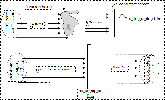
        [Please note that the radiographic film moves from the end position (in case of neutron exposure) to the centre position (in case of densitometer).]

        Speaker: Usman Kurshid Chaudhry (Non-Destructive Testing Group, PINSTECH, PAKISTAN)
      • 17:30
        Development and characterization of a neutron tomography system for the research reactor 1h 30m
        Waleed Abd el Bar (1) , Imbaby I. Mahmoud (2) , Hussein A. Konber (3) 1) Atomic Energy Authority (AEA), ETRR-2 . P. O. Box 13975, Abu Zabal, Egypt 2) Atomic Energy Authority(AEA), Research Centre, Engineering Department, Inshas, Cairo 11511 Egypt 3) Al Azhar University, Electrical Engineering Department, Nasr City, Cairo 81624 Egypt. Email:Engwaleed84@yahoo.com. Neutron tomography is a very powerful technique for the non-destructive evaluation of heavy industrial components as well as for soft hydrogenous materials enclosed in heavy metals, which are usually difficult to image using X-rays. It has found a variety of applications in medicine, agriculture and other heavy industries. In our effort to use this technique for non-destructive testing, a process has begun to upgrade the neutron radiography facility from static-based film (Nitrocellulose film and Agfa Structurix D7photographic film) neutron radiography into a dynamic neutron radiography/tomography system by using scintillation screens (ZnS(Ag)-6LiF) and a CCD-camera. Several experiments have been performed on this experimental station to study the feasibility of neutron tomography for various applications.
        Speaker: Abd el Bar Waleed (Atomic Energy Authority. Egypt)
      • 17:30
        Enhancement the safety of the Jordan research and training reactor (JRTR) 1h 30m
        Khalifeh AbuSaleem (1,2) 1) Jordan Atomic Energy Commission (JAEC) P.O.Box 70 Amman 11934- Jordan 2) Department of Physics The University of Jordan Amman 11942- Jordan Email: khalifeh.AbuSaleem@JAEC.GOV.JO The JRTR is a multipurpose reactor designed and constructed to be used for education and training, research and radioisotope production. All safety aspects of the JRTR fall under the category of SC-3 according to the ANSI/ANS 51.1 classification system of nuclear reactors. For example, the Reactor Structure Assembly (RSA), Primary Cooling System (PCS), CRDM/SSDM, Reactor Protection System (RPS), Confinement Isolation Dampers, Siphon Breaking Valves and UPS are classified as SC-3 components. However, in the wake of Fukoshima-Daicci accident, and learning the lessons of the accident and following the recommendations, the safety measures of the JRTR have been extensively investigated to enhance the safety of the reactor. Therefore, design changes of systems and equipment due to the reinforced international safety norm after Fukushima disaster, addition, expansion and modification of facilities to accommodate the design changes have been implemented. As a result investigation, several aspects of the JRTR safety have been improved. As examples of these, the quality class has been upgraded for several components such as Process Instrumentation and Control System (PICS), Radiation Monitoring System (RMS), Information processing System (IPS) and Operator WorkStation (OWS). Additionally, expansion and modification of facilities to accommodate systems and equipment have been applied. The seismic monitoring system has been improved by upgrading quality class and by adding a function generating the automatic seismic trip signal when a seismic motion exceeds Operating Basis Earthquake (OBE). Pool Liner Integrity has been enhanced by improving the welder qualification process and by enhancing the weld quality. Furthermore, the emergency conditions have attracted special attention. The emergency water storage capacity has been increased, and two mobile diesel generators have been placed in a building of seismic category I. This paper describes the safety aspects of the JRTR and the improvements after the Fukoshima-Daicci accident.
        Speaker: AbuSaleem Khalifeh (JAED and The University of Jordan, Department of Physics)
      • 17:30
        Feasibility analysis for the extraction of a thermal NR beam at the MNSR reactor 1h 30m
        M.H. Choopan Dastjerdi, J. Khorsandi, J. Mokhtari, A. Asgari Nuclear Science and Technology Research Institute, Tehran, Iran, Postal Code: 1439951113 Email: mdastjerdi@aeoi.org.ir In order to expanding the utilization of MNSR reactor, the possibility of extracting an appropriate thermal neutron beam for neutron radiography (NR) application is investigated. According to the physical restrictions of the MNSR, neutron beams are designed based on the vertical-tangential and oblique-tangential directions. Also, a thermal column is considered to reduce energy of neutrons. All designs are done by considering the least possible changes in the current reactor status. Results show that it is possible to obtain an appropriate NR beam with thermal neutron flux of about $ 2.53 × 10^6 n.cm^{-2}.s^{-1}$. The diameter and the collimation ratio of the obtained neutron beam at the image plane are 24 cm and 96, respectively. In addition, the thermal neutron flux has a good uniformity at this plane (flux fluctuation is <5%).
        Speaker: Dr Mohammad Hossein Choopan Dastjerdi (Nuclear Science and Technology Research Institute)
      • 17:30
        First attempts to use the dynamic neutron imaging method 1h 30m
        Marin Dinca Institute for Nuclear Research, Campului Str., No. 1, POB 78, 115400 Mioveni, Arges County, Romania Email: marin.dinca@nuclear.ro The presentation shows the preparation, started this year, for the imaging facility (INUS) placed at the tangential channel of the TRIGA Annular Core Pulsing Reactor from the Institute for Nuclear Research (INR) to achieve dynamic (real-time) imaging for applications in industrial field. This new method in INR involves EM-CCD Hamamatsu C9100-02 camera using new lens (F number 0.95) with better gathering of the light emitted by scintillators and an improvement in neutron intensity, with a better transfer of thermal neutrons from reactor core to collimator. The aim is the application of the method to industrial field for testing the behaviour of complex moving mechanisms that contain solids and liquids. In the near future it is intended to have INUS an instrument available for research and development through tomography and real-time imaging as non-destructive examinations for requests coming from research institutes and industry.
        Speaker: Dr MARIN DINCA (Institute for Nuclear Research)
      • 17:30
        Implementation of a new and high quality neutron radiography beamline at the Tehran research reactor 1h 30m
        M.H. Choopan Dastjerdi, H. Khalafi, Y. Kasesaz, A. Movafeghi Nuclear Science and Technology Research Institute, Tehran, Iran, Postal Code: 1439951113 Email: mdastjerdi@aeoi.org.ir A new neutron collimator as an important part of a neutron imaging facility is designed, installed and experimentally characterized at the Tehran Research Reactor. The design calculations are performed using MCNP monte Carlo code. Preliminary experimental characterization of the beam shows a thermal neutron flux of about $ 6.1×10^6 n\ cm^{-2} s^{-1}$ and a N/G ratio of about $ 4.82×10^5\, n\,cm^{-2} mrem ^{-1}$ at the 3-m image plane (L/D=150). Furthermore, the obtained neutron beam is characterized using the ASTM BPI and SI indicators and measurements indicate that the obtained radiographic image at this beam is of Category-I beam quality as defined in ASTM E545 standard.
        Speaker: Dr Mohammad Hossein Choopan Dastjerdi (Nuclear Science and Technology Research Institute)
      • 17:30
        Neutron imaging researches and applications in Brazilian research reactors: challenges and trends 1h 30m
        Ailton Fernando Dias and Altair Souza de Assis, Comissao Nacional de Energia Nuclear (CNEN), Brazil Email: ailton.dias@cnen.gov.br In Brazil, we have three research reactors with neutron extractor devices: 1) IEA-R1, at the Nuclear and Energetic Research Institute, in São Paulo; 2) IPR-R1, at the Nuclear Technology Development Centre, in Belo Horizonte; and 3) Argonauta, at the Nuclear Engineering Institute, in Rio de Janeiro. These facilities have been used for neutron imaging for more than 40 years, resulting in several technical reports, scientific papers, M.Sc. dissertations and Ph.D. thesis. In most of the cases, neutron imaging was used for testing and analyses of different kind of materials. At IEA-R1 Research Reactor, neutron imaging is an important research area, by using direct conversion (gadolinium plates) as indirect conversion (dysprosium plates). Neutron flow features of IEA-R1 at 2MW are: thermal flux of $3 × 10⁶ n/cm^2.s$; thermal to epithermal ratio of 5:7, cadmium rate of 150; n/γ ratio of $5 × 10^5 n/cm^2.mrem$; beam diameter of 20 cm. Neutron imaging has been used here to materials inspection and testing. Recently, real neutron imaging experiments have been carried out for inspection of samples in motion. At Argonauta Research Reactor, neutron imaging radiography has been carried out since 1972. Nowadays, Electronic Imaging Systems (EIS) offer real time inspection of samples, allowing dynamic events observation as well the inspection of a great number of samples per time unit. At 360W power, the characteristics of the Argonauta neutron facility are: thermal flux of $4,46 × 10^5 n/cm^2.s$; L/D ratio of 70; n/γ ratio of $3 × 10^6 n/cm^2.mrem$; average energy of 30 MeV. The real time neutron imaging applications includes drugs and explosives identification and localization, as well as materials testing for aircraft industry. At IPR-R1 Research Reactor, the first studies on neutron extractor installation were developed in 70´s, but only in 1987 this device was assembled and tested. It was used a vertical extractor, where samples were mounted in a box containing a photographic film and a gadolinium plate, placed at 3,62m from the reactor core, under a thermal flux of $1,68 × 10^6 n/cm^2.s$ (at 100kW power) and beam diameter of 10cm. This facility was used to examine several objects of different materials. Images obtained by neutron imaging were compared to X-ray imaging, as a way to identify in which cases we could use neutron or X-ray imaging. At this moment, a multipurpose reactor is under construction in Brazil, with expectation to be in operation before 2023. It will offer 13 neutron beams, where 7 of them will be in thermal neutron spectrum with energies between 10 to 100 MeV and 6 beams in cold neutrons spectrum with energies lower than 10 MeV. This new reactor will have a neutron extractor device with only one thermal neutron beam dedicated to neutron imaging. These thermal neutrons will be collected directly from the reflector tank by optimized neutrons extractors to assure the desired amplification factor at the samples position. In order to face the new challenges of radiopharmaceutical production and material testing as well as neutron activation and neutrongraphy, the operation of the multipurpose reactor will offer a higher thermal neutron flux and physical installations specially designed for neutron imaging. This new facility and the experience acquired by more than 40 years of neutron imaging researches will improve the Brazilian capabilities in development of new research areas, applications and materials testing by neutron imaging.
        Speaker: Ailton Fernando Dias (CNEN, Brazil)
      • 17:30
        Neutron radiography for cultural heritage objects in Iran 1h 30m
        A. Movafeghi(1), E. Yahaghi (2), M. H. Choopan-Dastjerdi (1) and B.Rokrok (1) (1) Nuclear Science and Technology Research Institute, Tehran, Iran (2 )Imam Khomeini International University, Qazvin, Iran Email addresses: amovafeghi@aeoi.org.ir Keywords: Neutron radiography; Cultural heritage; Non-destructive testing Neutron radiography (NR) is a useful technique in non-destructive testing (NDT) of cultural heritage objects. NR is complementary to X and gamma radiography. A new NR beam line has recently been built at the Tehran Research Reactor (TRR) in order to expand the national applications of NR. The examination and characterization of internal structure and composition can be difficult task, in particular for cultural heritage objects. Conventional Radiography shows only density variations and has some other drawback in archaeological applications, particularly as the more durable metals such as gold, silver and lead are nearly opaque to X-rays. On the other hand, NR is quite sensitive in detection of hydrogenous materials such as water, water-logged ceramics, organic materials such as wood or water-logged wood, plants, seeds, food remnants, leather, textiles, paper, fragrances, tar, epoxy resins etc. With NR, for example, it is possible to visualize hydrogen-containing materials inside metal artefacts much better than with X-rays, whereas the XR technique is more suitable for visualizing the integrity of metal objects or metal parts of objects made of organic materials. In this research, a historical object has been radiographed by means of new neutron beam line of TRR. The object was a vase from Samiran region of Qazvin Province, Iran. The age of the vase has been estimated 900 years old, approximately. The digital neutron radiography technique was used by the digital imaging plates (or Computed Radiography: CR). The image was obtained and saved in digital format. This was the first neutron radiography image of new NR facility at TRR. The results showed that the new system can be used effectively for the neutron radiography of cultural heritage objects.
        Speaker: Amir Movafeghi (Nuclear Science and Technology Research Institute, Tehran, Iran)
      • 17:30
        Neutron radiography of hydrogen redistribution in Zircaloy 1h 30m
        Weijia Gong (1), Pavel Trtik (2), Johannes Bertsch (1) 1) Laboratory for Nuclear Materials, Paul Scherrer Institut (PSI), Switzerland 2 )Laboratory for Neutron Scattering and Imaging, Paul Scherrer Institut (PSI), Switzerland Email: weijia.gong@psi.ch During the operation in a nuclear reactor, zirconium cladding tubes take up a part of the hydrogen produced by the oxidation reaction between the reactor coolant water and the hot rod surface. This hydrogen is source of degradation mechanisms affecting cladding safe operation under normal and accidental conditions. Provided the general tendency for hydrogen to diffuse down thermal and concentration gradients, and up stress gradients, hydrogen distribution can often be non-uniform within components, which could arise the risk for the integrity during intermediate dry storage, handling and transportation. Neutron radiography provides a powerful approach to investigate this non-uniform field of hydrogen concentration, given that the absorption cross-section of hydrogen for neutron is about one order of magnitude higher than that of zirconium. A most typical case of hydrogen redistribution is delayed hydride cracking (DHC) where hydrogen diffuses along the elevated stress to the crack tip area followed by hydrides formation and cracking. In order to look into this hydrogen diffusion process driven by stress, a meso-scale 3-point bending equipment that has been constructed in our group especially for in-situ neutron imaging, was tested in an ex-situ way at the BOA beamline (SINQ spallation neutron source, PSI) using PSI ‘Neutron Microscope’ instrument [1]. The cold energy spectrum at BOA allows for high hydrogen sensitivity. A notched Zircaloy-4 sheet was pre-charged with ~ 600 wppm of hydrogen followed by annealing for homogeneity. Then the sample underwent a thermo-mechanical cycle of DHC test. Before and after the test, neutron imaging was consecutively performed to determine the variation of the H concentration field due to the stress. The results revealed an area of hydrogen concentration at the notch, with a concentration gradient detectable over 700 $\mu$m. A maximum elevation of 160 wppm hydrogen was found at ~200 $\mu$m off the notch, where the largest tensile stress is located according to the FEM analysis. The measurement of neutron radiography is in very good agreement with our modeling result yielding an elevation value of 250 wppm of hydrogen concentration. Another interesting case concerns liner claddings which have been widely used in Swiss LWRs, either an inner liner to protect from PCI in BWRs or an outer liner (DX-D4) to improve corrosion resistance in PWRs. In post-irradiation examination, one always observes high hydrogen contents in liner material, visible as high concentration of hydrides, and a depleted hydrides density in the nearby cladding area. In the previous study, we have assessed qualitatively the effect of different liners on the distribution of hydrides in cladding cooled down at two different cooling rates. However, the quantification of hydrogen concentration in the liner still remains in mystery as well as the gradient at the liner/matrix interface, because the conventional measurement of hot gas extraction provides no spatial information. With this context, the determination of hydrogen concentration across various liner claddings is to be performed in our SINQ beam campaign scheduled in July 6th-8th. We would like to present these fresh results at the IAEA training workshop. Keywords: hydrogen, diffusion, stress, neutron radiography. [1] P. Trtik, E. H. Lehmann. Progress in High-resolution Neutron Imaging at the Paul Scherrer Institut - The Neutron Microscope Project. Journal of Physics: Conference Series 2016;746:012004.
        Speaker: Dr Weijia Gong (Paul Scherrer Institut)
      • 17:30
        Neutron radiography: Research, application and recent developments in Bangladesh 1h 30m
        S. Saha (1), M. N. Islam (2), B. Schillinger (3) 1) Institute of Nuclear Science and Technology, Bangladesh Atomic Energy Commission 2) Training Institute, Bangladesh Atomic Energy Commission 3) Heinz Maier-Leibnitz Zentrum (FRM II), Technische Universitaet Muenchen, Germany Email: sudipta.sust@yahoo.com The 3 MW TRIGA Mark II research reactor at Atomic Energy Research Establishment (AERE) in Dhaka, Bangladesh, was commissioned in 1987. At the time, a neutron radiography facility for film was installed. In these days, the facility has been utilized to support the innovations of different laboratories inside and outside of Bangladesh Atomic Energy Commission. Various types of materials including hydrogel/super water absorbent, bandages made of collagen, polymer, conventional and unconventional bricks have been taken under investigation independently for quality control and water absorption characteristics study. The facility is providing support to investigate coral samples from St. Martin islands for the blue economy program of People’s Republic of Bangladesh government. In addition to it, the facility is taking part into fabrication of high efficiency neutron shielding materials. Nevertheless the digital neutron radiography machine installation is still in progress.
        Speaker: Sudipta Saha (Institute of Nuclear Science and Technology)
      • 17:30
        Observation of hidden archaeologic relics using neutron radiography 1h 30m
        Jin Man Kim(1), TaeJoo Kim(1), JongYeul Kim (1) 1) Neutron Science Center, Korea Atomic Energy Research Institute, Daejeon, South Korea Email: kjm5@kaeri.re.kr Cultural heritage is ours from the past and should be passed on to future generations. They contain a unique and irreplaceable historical record for understanding the history. Interestingly, some heritages include hidden relics inside them. Many researchers have tried to analyze information about the hidden relics to complete a preserved historical puzzle without any damage to the relics, which may occur during observation. In this respect, non-destructive testing technology (NDT) is very important for the inspection of archaeological objects. Of the many available NDT, X-ray radiography has been widely used and should now be routine for research and conservation of most archaeological objects and other relics. A complementary neutron radiography is much less used in this area. Until recently, the main reason for this was the very small availability of moderately easily accessible neutron sources and the lack of efficient modern neutron imaging equipment. However, Rant and Kardjilov [1] explored the advantages of neutron which can penetrate metals. Also, Schillinger [2] reported that neutron NDT (NNDT), i.e. tomographic scanning, enables to disclose hidden characteristics for relics. Neutron radiography improves the knowledge and understanding about the past cultures. In specific, tomographic scanning is a powerful method for studying of archaeological objects. Therefore, the various characteristics of the object can be examined by neutron radiography. References [1] J.J. Rant, in: The Eighth International Conference of the Slovenian Society for Non-Destructive Testing, Portorož, Slovenia, 1–3 September 2005, pp. 181–188. [2] B. Schillinger, et al., in: Proceedings of Fifth World Conference on Neutron Radiography, Berlin, Germany, 17–20 June, 1996.
        Speaker: Jan Min Kim (Neutron Science Centre, Korea Atomic Energy Reserach Institute, Daejon, South Korea)
      • 17:30
        Primary experiments and inspections of materials using new NR beamline at the TRR 1h 30m
        M.H. Choopan Dastjerdi, A. Movafeghi, B. Rokrok, H. Khalafi Nuclear Science and Technology Research Institute, Tehran, Iran, Postal Code: 1439951113 Email: mdastjerdi@aeoi.org.ir Neutron radiography is a powerful technique for non-destructive testing of materials forindustrial application and research. This technique is complementary to X-ray radiography and finds unique applications in quality assurance of nuclear fuel and investigation of cultural heritage objects. Anew neutron radiography beam line has recently been built at Tehran Research Reactor in order to expandthe application of neutron radiography. In this work, some experiments and inspections of various samples like fuel rods, a cultural heritage sample and some flowers are done. In the qualitative investigations of fuel rods, the difference in size and enrichment between the pellets and the gaps between them were obviously recognized in the neutron radiographic image. In the quantitative investigations, data of the pellets compositions, their sizes (lengths and diameters) and the gaps between them were extracted from obtained images. In the neutron radiographic image of the rose, the structure of the pedicel and the layered petals are well visible.
        Speaker: Dr Mohammad Hossein Choopan Dastjerdi (Nuclear Science and Technology Research Institute)
      • 17:30
        Quality assessment of the radial and tangential NR beamlines of the TRR 1h 30m
        M.H. Choopan Dastjerdi, A. Movafeghi, H. Khalafi, B. Rokrok Nuclear Science and Technology Research Institute, Tehran, Iran, Postal Code: 1439951113 Email: mdastjerdi@aeoi.org.ir To achieve a quality neutron radiographic image in a relatively short exposure time, the neutron radiography beam must be of good quality and relatively high neutron flux. Characterization of a neutron radiography beam, such as determination of the image quality and the neutron flux, is vital for producing quality radiographic images and also provides a means to compare the quality of different neutron radiography facilities. This paper provides a characterization of the radial and tangential neutron radiography beamlines at the Tehran research reactor. This work includes determination of the facilities category according to the American Society for Testing and Materials (ASTM) standards, and also uses gold foils to determine the neutron beam flux. The radial neutron beam is a Category I neutron radiography facility, the highest possible quality level according to the ASTM. The tangential beam is a Category IV neutron radiography facility. Gold foil activation experiments show that the measured neutron flux for radial beamline with length-to-diameter ratio $ (L / D) =150$ is $6.1 × 10^6 n cm^{-2} s^{-1}$ and for tangential beamline with $ (L / D) = 115$ is $2.4 × 10^4 n cm^{-2} s^{-1}$.
        Speaker: Dr Mohammad Hossein Choopan Dastjerdi (Nuclear Science and Technology Research Institute)
      • 17:30
        Software development for neutron computed tomography at Thai research reactor (TRR-1/M1) 1h 30m
        C. Tippayakul, J. Channuie, S. Wonglee, R. Picha, S. Khaweerat Thailand Institute of Nuclear Technology, Ongkharak, Nakorn Nayok, 26120 Thailand Email: jatechanc@tint.or.th During the last couple of years, the manual control of data acquisition for neutron computed tomography at a 1.2-MW TRIGA Mark III reactor, Thai Research Reactor (TRR-1/M1), were difficultly operated. A simple system for data acquisition with control software for the newly renovated neutron tomography facility has been developed using LabVIEW. The hardware of the system consists of a programmable CCD camera combined with a static stepping motor. The software was in-house developed to replace the previous one which is no longer used due to its capacity limitations. The new software is capable of displaying live images and automatically recording the images on a computer. In order to obtain optimal image quality, the software drives the image capture processes by adjusting camera temperature, exposure time and number of projections as well as images integration in certain frame numbers. For the neutron tomography setup, the software takes particular snapshots automatically at a sample position in line with the stepping movement of the rotating sample holder. Subsequently, the snapshots were saved in picture and numerical formats for further image processing. The new controller software has successfully tested for automatic real time data acquisition providing the appropriate input for tomographic reconstruction. The success in development of controller software contributes to the productivity and safety of neutron imaging routine at TRR-1/M1.
        Speaker: Mr Jatechan Channuie (Thailand Institute of Nuclear Technology)
      • 17:30
        Spatial resolution study of a neutron imaging system using the slanted edge method 1h 30m
        L. Boukerdja, O. Dendene, A. Ali Nuclear Research Center of Birine, Algeria Email: boukerdjal@yahoo.fr Neutron imaging is a very powerful technique for nondestructive testing; it allows obtaining image of the internal structure of objects in 2D or 3D mode. During the last years, attempts have been made to implement and to develop a neutron tomography system at the Nuclear Research Center of Birine. At this end a new Peltier-Cooled CCD (16 bit) has been installed instead the old CCD camera (8 bit) and a turntable has been designed. The aim of this work is to calculate the spatial resolution of a CCD camera and a scintillator based neutron imaging system by the measurement of the MTF which is obtained by a slanted edged image. On the other hand, we will give some results obtained through the characterization of the image detector such as the variation of the gray level as a function of exposure time and the effect of the cooling of the CCD camera on the noise signal.
        Speaker: Layachi Boukerdja (Centre de Recherche Nucléaire de Birine (CRNB), Algeria)
      • 17:30
        Status of the Jordanian research and training reactor (JRTR) 1h 30m
        Khalifeh AbuSaleem (1,2) 1) Jordan Atomic Energy Commission (JAEC) P.O.Box 70 Amman 11934- Jordan 2) Department of Physics The University of Jordan Amman 11942- Jordan Email: khalifeh.AbuSaleem@JAEC.GOV.JO The JRTR has been built to be the corner stone of the center for excellence in nuclear sciences and technology. It is a multipurpose, $\text 5\ MW_{th}$ upgradable to $\text10\ MW_{th}$ research reactor. It uses the well proven LEU fuel plates of $U_3Si_2$ in Aluminum matrix. The thermal neutron flux in the core center has been measured to $1.7 x10^{14} / cm^2s$. The JRTR is equipped with 22 in-core irradiation locations mainly for Neutron Transmutation Doping (NTD), Neutron Activation Analysis (NAA), Radioisotope Production (RI), Research, and Training and Education. Particularly, three facilities for $^{192}\text Ir$, $^{131}\text I$, and $^{99}\text Mo$ production using neutron activation will be operational at the startup date. Also On-Power Loading and Unloading (OPLU) of targets shall be possible for $^{192}\text Ir$ and $^{99}\text Mo$. Three facilities, low-$\gamma$ environment, for NAA will be operational. In particular, NAA requires thermal and epithermal spectra; two thermal and one epithermal spots will be available. The NAA Facility is equipped with three Pneumatic Transfer Systems (PTSs). Very soft spectra can be utilized (future plan). Two NTD locations for up to 6-inch ingots and one for up to 8 inches are available. Three other locations for future expansions will be available. For the ex-core irradiation services, there are four tangentional beam ports and a one thermal neutron column facing the core. The beam ports will be used for Neutron Radiography (NR), and two beam ports for Standard Applications (ST). In addition, a future Cold Neutron Source (CNS) is planned to be installed. The initial criticality has been achieved using external neutron source on April 25, 2016, and all planned hot commissioning tests have been carried out successfully. The planned Neutron Radiography Facility (NRF) at JRTR will be at the level of the state-of-the-art facilities worldwide. It will cover all aspects of standard applications like neutron radiography and tomography. The instrument can accommodate large spectrum of research projects related to cultural heritage, materials science, energy renewable sources, geology, biology and fundamental science. Innovative experimental methods like phase-contrast imaging and real-time radioscopy will be possible. Industrial applications can be performed with a great success at this instrument. The facility will be instrumental for serving the needs of the future neutron and X-ray imaging community in Jordan and in the region.
        Speaker: AbuSaleem Khalifeh (JAEC and The University of Jordan, Dep. of Physics)
      • 17:30
        Study of reactor structural materials at the neutron imaging beam line Dhruva, India 1h 30m
        Shefali Shukla, Yogesh Kashyap, Tushar Roy, Mayank Shukla and S.C.Gadkari Technical Physics Division, Bhabha Atomic Research Centre, Mumbai – 400085, Maharashtra, India Email: shefali@barc.gov.in Reactor sources are best suited for advanced imaging techniques such as tomography and phase contrast imaging applications because they have large and stable thermal neutron flux. A neutron imaging beamline has been designed and developed at HS-3018 port of Dhruva reactor (100MW research reactor) and is currently being used for thermal neutron tomography and phase contrast imaging applications. This paper discusses about the experiments which have been done till date on this facility. A brief overview of the beamline design is also provided. The collimator has been designed in such a way that tomography or phase contrast imaging studies can be performed on the same beamline. A sapphire crystal as neutron filter followed by a bismuth crystal for gamma filtering has been used at the input of the collimator to maximize the neutron-to-gamma ratio. The maximum beam size of neutrons has been restricted to ~ 140 mm diameter at the sample position. A cadmium ratio of ~ 250 with L/D ratio of 160 and thermal neutron flux of $~ 4x10^7 n /cm^2 / s$ at the sample position has been measured. Study of hydrogen in reactor materials is particularly important to prevent the hydride induced embrittlement. In many cases, what we need information within bulk of material. Neutrons play a major role as they can penetrate through dense materials with added benefit that one can analyze the sample non- destructively. We have studied hydrogen ingression in reactor clad materials and also quantified the amount of hydrogen present. Neutron tomography studies on blisters in Zr-alloy have been carried out as an aid to NDT. In addition to conventional tomography neutron phase imaging technique are also being explored for those samples which have low neutron absorption cross-section.
        Speaker: Mrs Shefali Shukla (Bhabha Atomic Research Centre)
      • 17:30
        The design of neutron imaging instrument combined with PGGA set up at Maamora Triga Reactor 1h 30m
        A. Ouardi Centre National de l’Energie, des Sciences et des Techniques Nucléaires (CNESTEN) Email: ouardi@cnesten.org.ma A new neutron imaging instrument will be built to support the area of neutron imaging research (neutron radiography and tomography At Maamora Triga research Reactor (CNESTEN Research Centre, Rabat). The instrument is designed for research community and for routine quality control for industrial, mining, automotive and aircraft applications. It will be also useful tool for assessing water damage in air craft components, and the study of archaeological artefact. This neutron imaging set up will be combined with Prompt gamma with the prompt gamma-ray activation. The whole system will be mounted on the tangential channel. Both techniques are complementary and their combination provides full picture about sample by obtaining the material’s composition and the spatial distribution of the material in the sample set up. In this configuration the convergent part consist on the association of material with capability to reduce rapid neutrons and gamma (Borate Iron, Borate polyethylene and lead), and a primary shutter. The installation of these parts is in process. The second part is housed in the Triga Reactor hall, and including: The drum exchanger Collimator, Flight tube and Beam delimiter. As defined in our previous works basing on Geant4 simulations, fast neutron (5cm sapphire) and gamma (5cm bismuth) filters will be inserted in the convergent part. The L/D drum exchanging is housing 4 pinhole collimator with apertures of 1cm, 2cm, and 2,5cm and will reduce the beam size to 8 cm x 8 cm, 12 cm x 12 cm and 20 cm x 20 cm at the detector position respectively. The whole instrument will operated in three different positions, one for high resolution and the other for high speed.
        Speaker: Dr Afaf Ouardi (Centre National de l'Energie des Sciences et des Techniques Nucléaires)
      • 17:30
        The implementation of a charge coupled device (ccd) camera in a neutron imaging system for real time and tomography investigation 1h 30m
        Khairiah Yazid, Muhammad Rawi Mohamed Zin, Rafhayudi Jamro, and Azraf Azman Malaysian Nuclear Agency Bangi 43000 Kajang Selangor MALAYSIA Email: khairiah@nuclearmalaysia.gov.my The Malaysian Nuclear Agency (Nuclear Malaysia) operates the one and only research reactor in Malaysia, Reactor TRIGA PUSPATI (RTP) of Mark II type, commissioned on 28 June 1982. It has a nominal power of 1 MW designed to effectively implement various fields of basic and applied nuclear research or services, education and training. The PUSPATI TRIGA is a swimming pool-type light water research reactor with enriched uranium-zirconium-hydride fuel and graphite reflector. There are three radial beam ports, one tangential beam port and one thermal column. The maximum steady state operating power of the reactor is 1MW and at this operating power the thermal neutron flux at the edge of the reactor core is around $\text 2.797 × 10^{12} n/cm^2 / sec$. Neutron radiography has been developed at one of the radial beam ports since 1980s, but with a stagnation of a long period. The direct exposure technique using Gadolinium foil converter and Kodak SR45 (SR5) film was established using this facility. However, this facility has low thermal neutron intensity at the sample position, which leads to long irradiation times; it gives many limitations for the industrial applications. A process has begun to upgrade the neutron radiography facility from film-based neutron radiography into digital neutron radiography. Now, the neutron radiography facility has been re-developed during these years, a new improved collimator has been planned and designed and a new instrument for neutron radiography and computed tomography will be set up at the neutron facility. A major step in the improvement of the neutron radiography activity at PUSPATI TRIGA Reactor is the implementation of digital neutron detector for fast neutron radiography. The new neutron detector is based on a scintillator, a front coated mirror, lenses and a cooled scientific CCD camera. Recently, preliminary testing after the implementation of digital neutron radiography based on CCD neutron camera has been done using SANS beam port due to neutron facility is currently under construction. The neutron beam intensity at SANS beam port is estimated to be $~ 10^3 n / cm^2 /$ s with the TRIGA reactor operating at 750kW. Several experiments have been performed on this experimental station using the new digital neutron CCD camera. The results have demonstrated that the new digital neutron CCD camera show high potential to inspect low-thickness samples. Until now just a few experiments were studied and a systematic study is still pending. More work will be explored on real time neutron radiography using the new digital neutron CCD camera at neutron facility beam port. The most important property, the performance of the imaging instruments will be quantified. As the conclusion, the research and development in neutron imaging by utilizing PUSPATI TRIGA reactor is positively active. Further support from IAEA and other member countries is needed especially related to the upgrading of the neutron radiography/tomography facility. We also wish to cooperate and exchange each other with all colleagues in the world!
      • 17:30
        Upgrading the neutron radiography set-up at IFE in Kjeller, Norway 1h 30m
        S. Deledda(1), C. Prabhu (1), G. Helgesen (1), H.K. Jenssen (2) 1) Physics Department, Institute for Energy Technology, PO Box 40, NO-2027 Kjeller, Norway 2) Department of Nuclear Materials Technology, Institute for Energy Technology, PO Box 40, NO-2027 Kjeller, Norway Email: stefano.deledda@ife.no The JEEP II research reactor (2 MW thermal power) at the Institute for Energy Technology (IFE) in Kjeller (Norway) is presently the only neutron source in the Nordic Countries. A national infrastructure project, funded by Research Council of Norway, to upgrade the neutron scattering facilities, NcNeutron, is currently running (2016-2020). NcNeutron aims at establishing a neutron research and technology exchange center and being a Norwegian and regional home-laboratory in neutron-based science. The upgrade of the facilities includes the reconstruction and modernization of the neutron radiography instrumentation which was built in the mid-1970s. The current setup is based on a traditional Dy-foil technique with a relatively high spatial resolution (40-50 µm), but with a low maximum number of recorded images per day and with a man-hour intensive image processing. The facility is presently dedicated to the analysis of post-irradiation examination data from safety and integrity tests of nuclear fuels performed within reactor safety research projects. The neutron radiography upgrade, currently in the design phase, consists of three main tasks: (i) beam optimization (new sapphire filter, new aperture, and new in-pile collimator); (ii) modification of the current radiography cell, implementing different configurations for the analysis of radioactive and non-radioactive samples; (iii) installation of new sample stage and new digital neutron imaging detector. The new neutron imaging instrument (NIMRA) is being designed to have an ratio of up to 300 and a FOV of $\text15\,x15\, cm^2$. It will be intended for studying energy materials (e.g. hydrogen storage systems, Li-ion batteries, heat storage units) as well as flow in porous media (e.g. clays, concrete). However, applications within other material classes and systems will be actively pursued.
        Speaker: Dr Stefano Deledda (Institute for Energy Technology)
    • 08:30 12:00
      Thursday
      • 08:30
        Applications of neutron imaging in research and industry 45m
        Michael Schulz, Heinz Maier-Leibnitz Zentrum (MLZ) michael.schulz@frm2.tum.de The field of applications of neutron imaging is very diverse and frequently new applications appear from the need of customers to visualize details in samples that could not be investigated with other non-destructive techniques such as X-ray CT. Due to their unique contrast mechanism, neutrons give particularly high sensitivity for hydrogen and good penetration of many metals. As a result, several types of investigations have been established as standard use cases for neutron imaging. An overview of such applications coming both from scientific and industrial background will be given in this presentation.
        Speaker: Michael Schulz
      • 09:15
        Bragg edge neutron imaging 45m
        Malgorzata Makowska, Heinz Maier-Leibnitz Zentrum (MLZ) Email: Malgorzata.Makowska@frm2.tum.de Energy resolved neutron imaging is a non-invasive technique based on spatially resolved measurements of the intensity of the neutron beam transmitted through the sample as a function of neutron energy / wavelength. When neutrons pass through a polycrystalline material so-called Bragg edges occur in the transmission or attenuation patterns. These features in the wavelength dependent attenuation spectra occur at the wavelength values corresponding to the lattice spacing, and thus can be used to achieve information about crystal structure, crystalline phases, texture or distortions of the lattice plains due to strain or temperature, that are present in a sample. Since Bragg edge patterns are obtained through measurements performed in an imaging geometry, the measured crystallographic information is provided with spatial resolution. Principles of the Bragg edge neutron imaging technique, required instrumentation and examples of applications will be presented.
        Speaker: Malgorzata Makowska
      • 10:00
        The ancient steel sword and armour technology revealed through advanced neutron imaging techniques 45m
        Francesco Grazzi Consiglio Nazionale delle Ricerche, Istituto dei Sistemi Complessi, Italy Email: francesco.grazzi@isc.cnr.it The metallurgy of historic arms and armours, is one of the most interesting topics in archaeometallurgy because these objects were manufactured, over the ages, using the highest quality materials and the most advanced technology [1-2]. In particular, the compositional and microstructural characterization of swords, particularly steel swords, can hence allow us to learn about the technological skill reached by different civilizations. The use of non-invasive techniques allows the study of museum objects in excellent conservation conditions thus giving a clear view of their characteristics, and neutron imaging is, to the authors’ knowledge, one of the best methods to study morphology, identifying non-metallic inclusions, cracks and defects [3-6]. Thanks to the use of advanced techniques, such as energy selective imaging, the microstructural features and the distribution of the different phases in steel can be determined [7-10] so gaining important information about composition and manufacturing treatments (both thermal and mechanical). Following this path, we have performed a number of experiments using neutron imaging to reveal the characteristics of many artifacts, from different civilizations, of which the production procedures are not yet fully clear. We have studied the complex structure and the thermal and mechanical treatments applied to produce Japanese swords, the microstructure of wootz steel used to produce the “watered silk” pattern on Indo Persian swords, the multilayered Fe-Ni alloys used to produce the Indonesian keris, composite renaissance swords from Solingen and Toledo, and pattern welded Viking swords. Concerning armours, we studied the method of making lamellar Japanese helmets and the microstructure of Indian armour pieces made of wootz steel. The results obtained non-invasively through neutron imaging allow us to identify unique features that can shed new light on the manufacturing methods thus increasing the level of our knowledge about the technological skill of such civilizations. References [1] A. Williams, The sword and the crucible, Brill, Leiden (2003) [2] V. F. Buchwald, Iron and steel in ancient times, Historisk-filosofiske Skrifter 29, The Royal Danish Academy of Sciences and Letters (2005) [3] E. Lehmann et al., Archaeometry 52, 416 (2010) [4] R. Triolo et al., Analytical Methods 6, 2390 (2014) [5] F. Salvemini et al., Eur. Phys. J. Plus 128, 87 (2013) [6] F. Salvemini et al., Appl. Phys. A 117, 1227 (2014) [7] F. Salvemini et al., J. Anal. At. Spectrom., 27, 1494 (2012) [8] S. Peetermans et al., Analyst 138, 5303 (2013) [9] E. Barzagli et al., Eur. Phys. J. Plus 129, 158 (2014) [10] F. Salvemini et al., Eur. Phys. J. Plus 129, 202 (2014)
        Speaker: Dr Francesco Grazzi (CNR-ISC)
      • 10:45
        Coffee 30m
      • 11:15
        IAEA e-learning tools 45m
        Nuno Pessoa-Barradas, International Atomic Energy Agency (IAEA) Email: N.Pessoa-Barradas@iaea.org Research reactors (RRs) have contributed for more than six decades and continue contributing to the advances in nuclear science and technology development in IAEA Member States (MS), including nuclear power. The sustainability of their life-cycle is an issue of major concern and MS are increasingly seeking Agency's assistance in addressing the main challenges related to RR sustainable operation, including effective utilization, as well as in building new and accessing existing RRs for developing their national nuclear programmes and strategies, including for development of human capital. There are currently 216 operational research reactors (RR) in the world, with another 41 planned, under construction or in temporary shutdown $^1$. The most widely used technique is Neutron Activation Analysis, with 118 RRs in 53 Member States. With many neutron activation analysis scientists and engineers retiring, and with many newcomers having a non-nuclear background, it behoves the national and international communities to pass on the information in a non-traditional format, namely e-learning. Since the middle 1990s, e-learning has slowly infiltrated and now integrates education at all levels. E-learning encompasses a wide array of deliveries from simple introductions of animations in a teaching class to Massive Open Online Courses (MOOCs) and Modular Object-Oriented Dynamic Learning Environment MOODLEs. E-learning takes an extra challenge where the subject matter is highly scientific and engineering oriented encompassing multiple complex mathematical notations and concepts. A straight forward presentation of equations in PowerPoint is a recipe for an unsuccessful delivery of lectures. The challenge exists of how to deliver these concepts without the use of a traditional blackboard. An e-learning tool for NAA has been developed by the IAEA for the Member States, to pass on the basics and more advanced forms of the technology in a manner such that both novices starting their careers in this area and higher level staff members and academics can utilize the modules developed to refresh themselves or to teach others. The modules are organised in seven categories covering all the essential aspects of NAA: Introduction, Basic Nuclear Physics, Instrumentation, Calibration, Quality, NAA Practice and Varieties of NAA. The e-learning tool for NAA will be presented, leading to a discussion on future expansion to other areas of utilization of RRs. There is currently a substantial need to develop strategies for RR effective utilization on a national, regional and international basis, given that a significant number of these facilities are ageing and not utilized to their full potential. Neutron imaging is the second most common technique reported in the IAEA RRDB, with 72 facilities in 39 Member States. With many users coming from areas as diverse as materials research, cultural heritage or environmental studies, the need and scope for developing an e-learning course for neutron imaging will be discussed. $^1$ IAEA Research Reactor Data Base https://nucleus.iaea.org/RRDB/RR/ReactorSearch.aspx
        Speaker: Nuno Pessoa-Barradas (IAEA)
    • 13:00 17:30
      Experiments II
    • 08:30 12:15
      Friday
      • 08:30
        Imaging with polarized neutrons 45m
        N. Kardjilov (1), A. Hilger (1), I. Manke (1), J. Banhart (1) 1) Helmholtz Centre Berlin, Hahn-Meitner Platz 1, 14109 Berlin, Germany Email: kardjilov@helmholtz-berlin.de The neutrons are able to pass through thick layers of matter (typically several centimeters), but are sensitive to magnetic fields due to their intrinsic magnetic moment. Therefore, in addition to the conventional attenuation contrast image, the magnetic field inside and around a sample can be visualized independently by detection of polarization changes in the transmitted beam [1]. This is based on the spatially resolved measurement of the cumulative precession angles of a collimated, polarized, monochromatic neutron beam that transmits a magnetic field [2]. Solid state polarizing benders can be used to polarize and analyze a monochromatic neutron beam. The configuration allows for quantitative polarimetric experiments, where the polarization vector of the magnetic field associated with a sample is measured in three orthogonal directions. By applying an iterative algorithm to the measured rotation angles, it is possible to reconstruct the flux density of the 3D magnetic field that produced them. Polarizing filters based on polarized $^3 He$ gas can be used for high resolution imaging of magnetic materials using polychromatic neutrons. Neutron depolarization imaging allows for observations of phase transitions between ferromagnetic and paramagnetic states in single crystals, allowing position-sensitive mapping of the Curie temperature [3]. Examples of investigation of various magnetic materials will be presented. REFERENCES [1] N. Kardjilov et al, Nature Physics 4, 399-403 (2008) [2] M. Dawson et al 2009 New J. Phys. 11 043013 [3] M. Schulz et al 2010 J. Phys.: Conf. Ser. 211 012025 KEY WORDS: Polarized neutrons, Magnetic materials, $^3\text He$ cells, solid state benders
        Speaker: Dr Nikolay Kardjilov (Helmholtz-Zentrum Berlin)
      • 09:15
        Neutron depolarization imaging 45m
        Marc Seifert Heinz Maier-Leibnitz Zentrum, Technical University of Munich, Germany Email: marc.seifert@frm2.tum.de The neutron depolarization imaging (NDI) technique is based on the combination of a neutron imaging beam line with a neutron polarization analysis setup. It enables the spatially resolved measurement of the influence of a sample’s magnetic field on the neutron polarization. As the spin of a neutron precesses in magnetic fields due to Larmor precession, ferromagnetic (FM) samples depolarize the polarized neutron beam. NDI therefore can for example reveal inhomogeneities in FM samples by measuring the Curie temperature across the sample. As neutrons easily penetrate common cryostats and pressure cells, the samples can be investigated under extreme conditions such as low temperatures and high pressures.
        Speaker: Marc Seifert
      • 10:00
        The neutron imaging facility ODIN at the European Spallation Source (ESS) 45m

        M. Lerche (1), M. Morgano (2), M. Strobl (2)and E. Calzada (1)

        1)Technical University of Munich, FRMII & Heinz Maier-Leibnitz Zentrum (MLZ), Germany

        2) Paul-Scherrer-Institut, SinQ, Switzerland

        Email: Michael.Lerche@frm2.tum.de

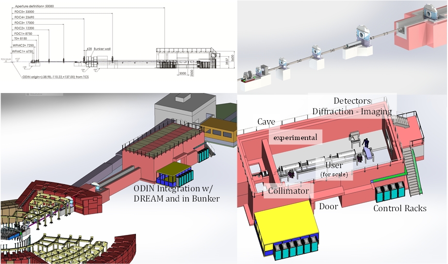
        ODIN (Optical and Diffraction Imaging with Neutrons) is a beamline project at the European Spallation Source (ESS). It is collaboration between the ESS, PSI and TUM, with TUM as lead institution.
        ODIN will provide a multi-purpose imaging capability with spatial resolutions down to the µm range. The pulsed nature of the ESS source will give access to wavelength-resolved information. Different imaging techniques, from traditional attenuation-based imaging to advanced dark field, polarized neutron or Bragg edge imaging, will be available within the full scope of ODIN with unprecedented efficiency and resolution. A summary of the technical full scope and its science application will be given and the updated conceptual instrument design including its challenges, see figure 1, will be presented.

        Image

        Speaker: Michael Lerche
      • 10:45
        Coffer 30m
      • 11:15
        Neutron imaging at Phoenix Nuclear Labs 30m
        Michael Taylor, Phoenix Nuclear Labs, USA michael.taylor@phoenixnuclearlabs.com Phoenix Nuclear Labs has designed, constructed and is currently testing a high-flux accelerator-based neutron generator to be used for thermal neutron imaging. The unit provides a source of moderated neutrons, collimated to an image plane where samples can be placed and various neutron detectors may be used to capture neutron images. The system is designed to yield $3 × 10^{11} n/s$, with an expected $1.4 × 10^4 n / cm^2-s$ thermal neutrons at the image plane at a given collimation ratio as predicted by Monte Carlo N-Particle (MCNP) transport code. The cadmium ratio, a metric to quantify how well thermalized the beam is, has also been modeled in MCNP. Current measurements of the above metrics are ongoing but appear to match design parameters well at the present. Neutron output is continually increasing by optimizing the accelerator performance. The system is expected to produce ASTM category III (or higher) neutron images based on the metrics of signal to noise ratio, contrast sensitivity, thermal neutron content, gamma content, scattered neutron contribution and image resolution. The current status of the system, in particular the imaging parameters, are discussed in this paper.
        Speaker: Mr Michael Taylor (Phoenix Nuclear Labs)
      • 11:45
        Final discussion 30m